Plethysmography measures change in volume — in venous work, how much the volume of a limb changes as blood fills and empties it — to assess how well the veins and their valves are working. Instead of imaging a vein, it treats the whole limb as a container and records how it swells and drains during defined manoeuvres, turning venous function into a set of volume and time measurements.
For a buyer, the defining point is that plethysmography is not ultrasound. It is a functional, volumetric test that sits alongside duplex imaging rather than replacing it: duplex shows which valve leaks and where, while plethysmography quantifies the overall haemodynamic effect on the limb. Understanding that split is the key to specifying and using it well.
The three main plethysmography methods
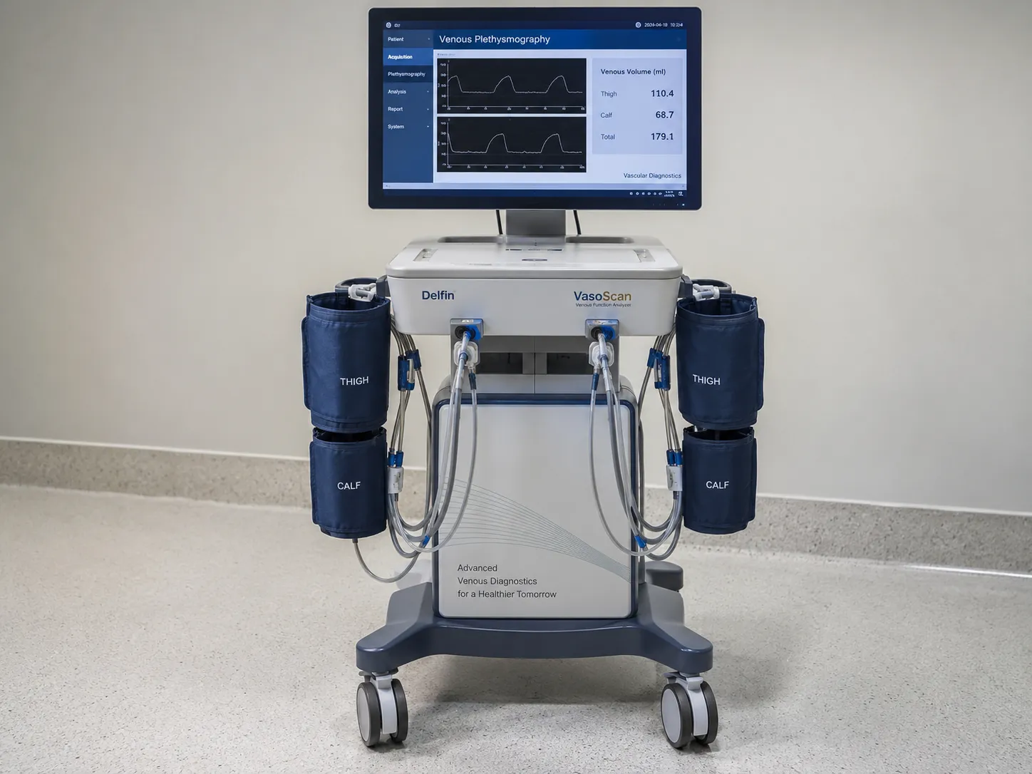
Several sensing principles carry the same name, and they are not interchangeable. Air plethysmography (APG) wraps an air-filled cuff or sleeve around the calf and measures volume change as the trapped air pressure changes, giving quantitative millilitre-scale figures. Strain-gauge plethysmography uses a fine mercury-in-silicone or indium-gallium gauge around the limb that stretches as the limb swells, changing its electrical resistance in proportion to circumference. Photoplethysmography (PPG) is different again: an optical sensor on the skin shines light and measures the reflected signal, tracking the volume of blood in the superficial tissue rather than the whole limb. Each answers a slightly different question, so match the method to the assessment you need.
Air plethysmography and the quantitative numbers
Air plethysmography is the method that yields the fullest set of venous figures. With the patient moving between lying, standing, and toe-raises, an APG system derives measurements such as venous filling index — how fast the calf fills with reflux — ejection fraction from a single toe-raise, and residual volume fraction after a series of them. Together these describe reflux severity, the strength of the calf muscle pump and how well the limb empties. Because the numbers are quantitative and repeatable, air plethysmography is used to grade venous disease and to assess the haemodynamic effect of treatment in a way an image alone cannot.
Photoplethysmography and venous refilling time
Photoplethysmography answers a narrower, simpler question and needs only a small optical probe. After the patient performs a set of ankle movements to empty the superficial veins, the PPG sensor tracks how quickly the blood refills the skin. A rapid venous refilling time indicates reflux; a normal, slower refill suggests competent valves. PPG is quick, inexpensive, and well suited to a screening or first-line role, though it is more qualitative than air plethysmography and reflects superficial rather than whole-limb volume. Many services use PPG as a fast triage and reserve APG or duplex for detailed assessment.
Where plethysmography fits alongside duplex
Duplex ultrasound is anatomical — it localises the failed valve — while plethysmography is functional, quantifying the overall effect on the limb. The two are complementary. A vein service may use duplex to map reflux and plethysmography to grade its haemodynamic severity or to measure calf-pump function before and after treatment. Plethysmography also has a role beyond veins: strain-gauge and related techniques feature in venous outflow and, historically, in deep vein thrombosis screening, though duplex has largely taken over there. Specify plethysmography for what it uniquely gives — quantified venous function — not as a cheaper substitute for imaging.
Standards, marking and electrical safety
A plethysmograph is medical electrical equipment built to the general standard IEC 60601-1, covering electrical safety and essential performance; systems that record physiological signals also fall under the collateral standards in that family. Because plethysmography is not ultrasound, the IEC 60601-2-37 ultrasonic standard does not apply to it — a useful check when a supplier bundles it with an imaging kit. The device must carry UKCA or CE marking, and device regulation and field-safety notices sit with the Medicines and Healthcare products Regulatory Agency at mhra.gov.uk. This guide draws on manufacturer documentation, the IEC 60601 family and UK regulatory guidance, and is written for UK procurement teams.
Calibration and why it decides the numbers
Plethysmography reports quantitative volumes, so calibration is not optional. Air plethysmography systems need their pressure-volume sensing calibrated on the manufacturer's schedule, and strain gauges must be sized and zeroed correctly for each limb or the resistance-to-volume conversion drifts. An uncalibrated plethysmograph produces plausible-looking numbers that are quietly wrong, which is worse than no measurement. Build periodic calibration and verification by your biomedical engineering team or the supplier into the running cost, and keep the certificates for audit, because every clinical figure the device reports depends on that calibration holding.
Consumables and the running cost
The sensing part is where the ongoing spend sits, and it varies sharply by method. Air plethysmography uses reusable air cuffs or sleeves that wear and need replacement. Strain-gauge systems use fine gauges that are delicate and have a limited life — mercury-in-silicone gauges in particular need careful handling and eventual replacement. Photoplethysmography probes and any single-use skin sensors are a recurring cost in a screening service. Budget for the sensor consumables, calibration, the service contract and software, and weigh them against test volume when you compare methods and suppliers.
Where plethysmography is used
Plethysmography turns up in vascular labs, vein clinics and some physiology departments. Air plethysmography suits a service that wants quantified venous function — grading reflux, measuring calf-pump performance and assessing treatment effect. Photoplethysmography suits primary-care-adjacent screening and quick first-line venous checks. Strain-gauge methods appear in venous outflow and research settings. Because the methods differ so much in output and cost, the right choice depends entirely on whether you need a quick qualitative screen or detailed quantitative haemodynamics, so define the clinical question before you choose the technique.
Reporting, records and connectivity
The output of a plethysmography study is a set of numbers and traces, so how they reach the record matters. Confirm the system stores and exports its venous filling index, ejection fraction, residual volume, or refilling-time results and the waveform traces into your reporting system, ideally electronically rather than as a paper printout alone. For a service tracking treatment effect over time, the ability to recall and compare prior studies is central, so check the export formats and how results are trended before you commit.
Beyond the purchase price
Costing a plethysmograph means looking past the console to the sensing and the calibration. The consumable sensors, the periodic calibration, the service contract, and any software licence all add up over the device life, and a strain-gauge system's fragile gauges can dominate the running cost. Weigh the upfront price against consumable and calibration spend and against how many studies you will actually run. When you compare plethysmography options and suppliers, use MediGear to compare suppliers on whole-life cost rather than headline price.
Outflow and occlusion plethysmography
Beyond reflux and pump function, plethysmography has an older role in measuring venous outflow. In occlusion plethysmography, a thigh cuff is inflated to block venous return while leaving arterial inflow; the limb swells to a plateau, and the rate at which it empties when the cuff is released reflects venous outflow and any obstruction. Strain-gauge and air methods both support this. Duplex has largely replaced plethysmography for diagnosing deep vein thrombosis directly, but outflow measurement still appears in venous obstruction assessment and research. If your service wants this capability, confirm the system offers a validated outflow protocol and the cuff hardware it needs, rather than assuming any plethysmograph covers it.
Specifying a plethysmograph: buyer's checklist
-
The right sensing method for your question — air plethysmography for quantitative haemodynamics, photoplethysmography for quick screening.
-
The full set of venous measurements you need, such as venous filling index, ejection fraction and residual volume.
-
A defined calibration and verification schedule, with certificates kept for audit.
-
Conformance to IEC 60601-1 and current UKCA or CE marking, with documentation supplied.
-
Sensor consumable costs — air cuffs, strain gauges or PPG probes — over the device life.
-
Electronic export of results and traces into your reporting system, with trend comparison.
-
Training so operators run the standing and toe-raise manoeuvres consistently.
The bottom line on plethysmography
Plethysmography measures the volume changes that reveal how well the venous system fills, empties and pumps — a functional test that complements, and never replaces, duplex imaging. Choose the method to fit the question, mind the calibration and the sensor consumables, and specify around IEC 60601-1 rather than the ultrasound standards that do not apply. If you are equipping a vascular or vein service, MediGear can help you compare verified suppliers and plethysmography options side by side.
Disclaimer
This article is for informational purposes only. It is published by MediGear (medigear.uk) for general information and procurement guidance, and is not clinical, diagnostic, treatment, technical, engineering, legal or regulatory advice, nor a product endorsement, guarantee or substitute for professional assessment. MediGear does not provide medical consultations. Buyers should consult their clinical, biomedical, estates and regulatory contacts, and the manufacturer's documentation, and independently verify all specifications, certifications, compatibility and suitability before purchase. Specifications, certifications and availability are correct at the time of publication and may change without notice. MediGear is a medical-equipment distributor and does not sell medicines or pharmaceutical products.