
A thoracoscope is an endoscope introduced through the chest wall to inspect the pleural space — the cavity between the lung and the chest wall — and to guide biopsy, fluid drainage and procedures on the pleura. It turns a blind space into a directly visualised one, letting a clinician see the parietal and visceral pleura, sample suspicious tissue, break down adhesions and place drains under vision. Thoracoscopy spans two worlds: physician-led medical thoracoscopy for pleural effusions and biopsy, and surgical video-assisted thoracoscopic surgery (VATS) in theatre.
For procurement the split matters. A pleural or respiratory service buying for medical thoracoscopy wants a straightforward, often rigid, sometimes semi-flexible system and reusable or single-use trocars. A surgical unit buying for VATS wants a full high-definition or 4K rigid stack with angled telescopes. The device is the same idea; the specification and cost differ.
How the pleural space is imaged
The pleural space is a thin potential cavity held under negative pressure, so it must be opened to be seen. In medical thoracoscopy the clinician creates a controlled pneumothorax or drains effusion fluid to separate lung from chest wall, then passes a trocar through an intercostal incision and introduces the thoracoscope. Illumination from a cold light source lights the cavity while the camera relays the view to a monitor. Biopsy forceps passed down or alongside the scope take parietal pleural samples under direct vision, and talc can be insufflated to achieve pleurodesis.
In VATS the same access principle is scaled up: one or more ports admit a rigid telescope and separate instruments, with one-lung ventilation collapsing the operative lung to open the hemithorax. The thoracoscope provides the magnified, illuminated view that lets the surgeon resect, staple and dissect without opening the chest. Whether physician-led or surgical, imaging depends on opening the space, lighting it and relaying a clear image.
Variants and how they compare
Rigid thoracoscopes
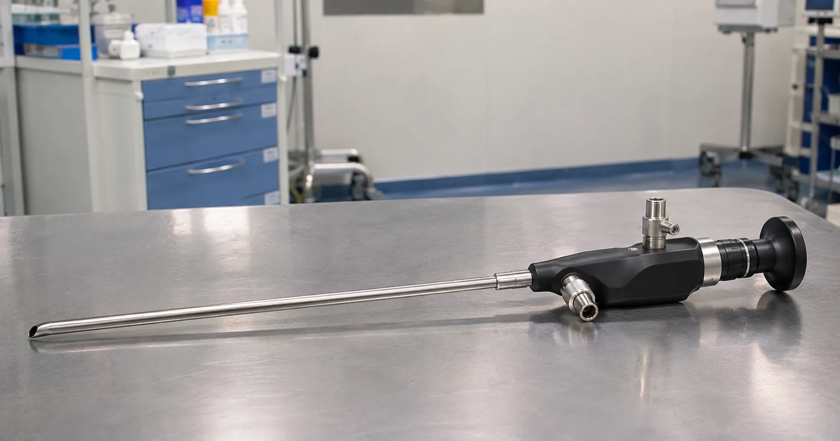
Rigid rod-lens telescopes give the brightest, sharpest image and are standard for both medical thoracoscopy and VATS. They come in angled views — commonly 0, 30 and 45 degrees — where an angled scope lets the operator see around structures without moving the port.
Semi-rigid and flexible thoracoscopes
Semi-rigid thoracoscopes, resembling a bronchoscope with a flexible tip on a rigid shaft, are popular for physician-led medical thoracoscopy because they are familiar to handle, integrate with existing endoscopy processors, and reach angled surfaces. The image is a little less crisp than a rigid rod lens, and the working channel constrains instrument size.
Single-use options
Single-use thoracoscopes and trocars remove reprocessing and cross-contamination concerns for lower-volume or infection-sensitive use, at a higher per-case cost.
Key specifications a buyer must check
For rigid systems check telescope diameter, working length and the range of viewing angles, plus the camera and processor resolution and end-to-end compatibility of camera, cable, light source and monitor. For semi-rigid scopes check the working-channel diameter, tip-deflection range and compatibility with your existing endoscopy stack and processor. A semi-rigid thoracoscope that shares your bronchoscopy processor avoids buying a second video tower, which can decide the economics for a physician-led service. Across all types confirm the trocar and port sizes, the biopsy and grasping instruments that fit, and whether the scope is autoclavable and sealed. Verify the light source is LED where possible for cooler running and long life. Consider whether you need talc-insufflation and pleural biopsy accessories, drainage catheters and, for VATS, the stapler and energy-device platforms your surgeons prefer, since these shape the instrument set as much as the scope itself.
Standards, regulation and documentation
A thoracoscope system is a medical device requiring valid UKCA or CE marking with instructions for use stating intended purpose and validated reprocessing. The camera console, light source and any electrosurgical or stapling energy device sit under the IEC 60601-1 electrical-safety family, with IEC 60601-1-2 for electromagnetic compatibility. Ask for the declaration of conformity and confirm Great Britain market registration. The MHRA regulates UK medical devices and receives safety reports, with broader guidance on GOV.UK and clinical guidance from NICE. Keep the instructions for use, which define the only validated cleaning and sterilisation route for the scope and channels.
Decontamination, calibration and maintenance
Reusable thoracoscopes enter a sterile field and must be sterile for each use. Rigid autoclavable telescopes are cleaned, inspected and steam-sterilised; semi-rigid scopes with a working channel need validated channel brushing, leak testing and high-level disinfection or sterilisation to their instructions for use, and camera heads and light cables follow their own rules. Optics need no clinical calibration, but cameras require white balance before each case and periodic image-quality checks. Inspect rod lenses for chips, fibres for delamination and channels for damage, since a degraded scope quietly erodes the view in a procedure where clear sight guides the biopsy and the drain.
Use across care settings
Medical thoracoscopy runs in respiratory and pleural services, often in an endoscopy suite or dedicated pleural procedure room, for investigating effusions and performing pleurodesis. VATS lives in cardiothoracic theatres for lung resection, pleural and mediastinal surgery. Single-use systems suit lower-volume units, isolation cases or services standing up a new pleural pathway without a reprocessing investment. Veterinary thoracoscopy exists on the same principles. Define which service you are equipping, because a physician-led pleural list and a VATS theatre need different stacks. A shared theatre stack can sometimes cover both if the endoscopy suite and theatre agree on a common camera platform, but reprocessing logistics and scope availability usually decide whether one stack or two is the better buy.
Consumables, spares and total cost of ownership
Recurring costs include trocars and ports, biopsy and grasping instruments, talc for pleurodesis, drainage catheters and, for surgery, staplers and energy devices, plus sterile drapes and camera covers. Rigid optics are the repair-sensitive item, so budget for repair or a service contract and hold a spare so a damaged scope never cancels a list. Ask about warranty on telescope, camera and light source separately, LED or lamp life, single-use scope pricing if you go disposable, and lead times on replacement cables and light guides. Our team can help you configure a medical-thoracoscopy or VATS stack to your caseload — register as a buyer or contact us to compare options.
Procurement checklist
-
Decide whether you are equipping physician-led medical thoracoscopy, surgical VATS, or both.
-
Choose rigid, semi-rigid or single-use scopes to match the service and reprocessing capacity.
-
Confirm viewing angles, telescope or channel diameter, and compatible biopsy instruments.
-
Check camera, processor, cable, light source and monitor resolution match end to end.
-
Verify the scope is autoclavable and sealed, or budget single-use per-case cost.
-
Request UKCA or CE documentation, IEC 60601 compliance and validated reprocessing instructions.
-
Agree warranty, spare cable and light-guide lead times, and loan cover during repair.
Conclusion
A thoracoscope opens the pleural space to direct view, guiding biopsy, drainage and surgery that would otherwise be done blind. The right buy depends on whether you run a physician-led pleural service or a VATS theatre, on rigid versus semi-rigid versus single-use, and on a reprocessing pathway you can resource. Match the angles, diameters and instruments to the work, confirm the paperwork, and cost the consumables and repairs honestly. MediGear can assemble a suitable stack so your pleural or thoracic team sees clearly and buys well.
Disclaimer
This article is for informational purposes only. It is published by MediGear (medigear.uk) for general information and procurement guidance, and is not clinical, diagnostic, treatment, technical, engineering, legal or regulatory advice, nor a product endorsement, guarantee or substitute for professional assessment. MediGear does not provide medical consultations. Buyers should consult their clinical, biomedical, estates and regulatory contacts, and the manufacturer's documentation, and independently verify all specifications, certifications, compatibility and suitability before purchase. Specifications, certifications and availability are correct at the time of publication and may change without notice. MediGear is a medical-equipment distributor and does not sell medicines or pharmaceutical products.