
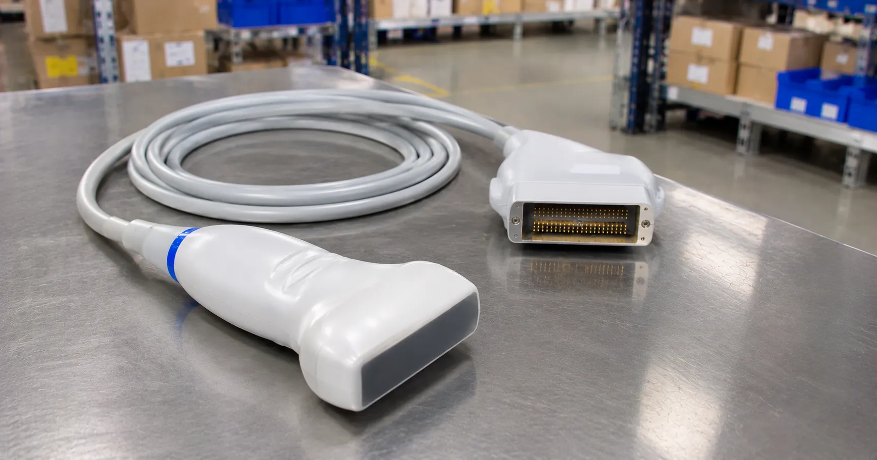
A Doppler ultrasound probe detects blood flow by sending sound waves into a vessel and measuring the frequency shift of the echoes that bounce back off moving red blood cells. Faster flow shifts the frequency more, so the device translates that shift into an audible signal or a waveform. For a procurement lead, the choices that matter are the probe frequency, whether the unit is continuous-wave or pulsed-wave, whether it is a simple handheld or a full duplex system, the display and connectivity, and the running cost of gel, batteries and probes over the life of the device.
The measurement principle
The Doppler effect describes how the frequency of a reflected wave changes when the reflector is moving relative to the source. A probe emits ultrasound at a fixed frequency; red blood cells moving towards the probe return a higher frequency, and cells moving away return a lower one. The device subtracts the two to give the Doppler shift, which falls in the audible range, so an operator can hear flow directly. The angle between the sound beam and the vessel affects the calculated velocity, which is why quantitative measurement needs a known insonation angle. In a simple handheld unit the output is sound; in a duplex scanner it is a colour map and a velocity waveform overlaid on a grey-scale image.
Continuous-wave versus pulsed-wave probes
Continuous-wave probes
A continuous-wave probe uses two crystals, one always transmitting and one always receiving. It detects high velocities without ambiguity but cannot tell you the depth the signal came from, because it listens along the whole beam. Handheld vascular and obstetric Dopplers are usually continuous-wave: cheap, rugged and ideal for a yes-or-no answer on whether flow is present.
Pulsed-wave probes
A pulsed-wave probe sends short bursts and times the returning echo, so it can sample flow at a chosen depth, the sample volume. This range resolution is essential for spectral analysis in a duplex study, but it limits the maximum velocity that can be measured without aliasing. Duplex and colour-flow systems combine pulsed-wave Doppler with imaging.
Probe frequency and what it means for buyers
Frequency sets the trade-off between penetration and resolution. Lower-frequency probes, around 2 to 5 MHz, penetrate deeper and suit deep vessels and larger patients. Higher-frequency probes, around 8 to 10 MHz, resolve superficial vessels well but do not reach as deep. Handheld vascular Dopplers commonly offer 8 MHz for peripheral arteries and 5 MHz for deeper or oedematous limbs, while obstetric probes use lower frequencies around 2 to 3 MHz for the foetal heart. Buying the right probe frequency for the intended vessels matters more than any other single choice, so match it to your caseload rather than defaulting to one size.
Sub-types across the range
At the simplest end are pocket handheld Dopplers with an audible output, used for pulse detection and the ankle-brachial pressure index. Next are handhelds with a small waveform display that show the flow trace, useful where the shape of the signal aids assessment. Full duplex and colour-flow scanners sit at the top: they image the vessel, map flow direction in colour and produce spectral velocity waveforms, but they cost far more and demand trained sonographers. Most primary-care, community and ward applications need only a handheld; vascular laboratories need duplex. Buy to the task, not the maximum capability.
Key specifications a buyer must check
Confirm the available probe frequencies and whether probes are interchangeable on one base unit, because a shared handpiece across 5 and 8 MHz probes saves money. Check battery type and life, or mains operation, and whether the unit is rechargeable. For units with a display, look at waveform clarity, the option to freeze and store traces, and any bidirectional capability that shows flow direction. Assess durability, water resistance for cleaning, and the quality of the audio output, which is what most users actually rely on. For duplex systems, the imaging specifications, transducer options and measurement software become the main comparison points.
Standards and UK regulation
A Doppler unit is an electrical medical device and must meet the IEC 60601-1 general safety standard, IEC 60601-1-2 for electromagnetic compatibility, and the particular standard IEC 60601-2-37 for ultrasonic diagnostic and monitoring equipment, which covers acoustic output safety. Any device on the UK market needs appropriate UKCA or CE conformity marking and, where required, manufacturer registration. Ask for the declaration of conformity and the acoustic output data. The MHRA publishes device safety information and field-safety notices for ultrasound equipment, and professional vascular and sonography bodies set the clinical protocols rather than the equipment vendor.
Calibration, maintenance and decontamination
Handheld Dopplers need little user calibration, but the probe and cable are the parts most likely to fail, so inspect them for cracks and lead damage that degrade the signal, and keep spares. Duplex systems need periodic performance checks and electrical-safety testing by biomedical engineering. Decontamination is a daily reality: probes contact skin and, for some applications, wounds, so follow the manufacturer's validated cleaning and disinfection method and use only compatible agents, because harsh chemicals crack the acoustic lens. Ultrasound gel is the standard coupling medium; single-use sachets reduce cross-contamination compared with refilled bottles, which is the safer choice in shared clinical areas.
Directional Doppler and connectivity
Some handhelds are bidirectional, separating flow towards and away from the probe into two channels, which helps distinguish arterial from venous signals and identify reversed flow. Simple non-directional units give a single audible signal only. If you document ankle-brachial pressure index results or waveforms in the patient record, confirm how the device captures and exports them, whether by printout, an on-screen trace you transcribe, or a digital output to software. A unit that stores and prints the waveform supports an auditable record, which matters where the result informs referral or wound management. For duplex systems, integration with the imaging archive and reporting system is a core requirement rather than an option.
Care settings, consumables and total cost of ownership
Handheld vascular Dopplers are used in GP surgeries, community nursing and leg-ulcer clinics for the ankle-brachial pressure index, on wards for pulse checks, in vascular access, and in veterinary practice. Obstetric handhelds are used in midwifery and antenatal clinics. Duplex scanners belong in vascular laboratories and imaging departments. The purchase price of a handheld is modest, so the lifetime cost is dominated by gel, batteries or charging, replacement probes and the occasional cable. For duplex systems the transducers, service contract, software updates and training dominate instead. Ask suppliers about warranty length, probe replacement cost, repair turnaround and loan availability. Standardising handheld models across a service simplifies training and spares. The MediGear buyers team can help compare options, or you can contact us to check availability and support.
Procurement checklist
-
Match probe frequency to the vessels you assess, and confirm probe interchangeability.
-
Decide between continuous-wave handheld and pulsed-wave duplex for your caseload.
-
Check battery life or mains operation, audio quality and any waveform display.
-
Confirm UKCA or CE marking, IEC 60601 compliance and acoustic output data.
-
Agree a validated probe cleaning method and compatible disinfectants.
-
Budget gel, batteries and replacement probes into total cost of ownership.
-
Confirm warranty, probe replacement cost and repair turnaround in writing.
Conclusion
A Doppler ultrasound probe turns the frequency shift of echoes off moving blood into a signal a clinician can hear or see, and the right choice depends on probe frequency, continuous- versus pulsed-wave operation and whether you need imaging at all. Match the device to the vessels and settings you actually work in, budget for probes and consumables, and confirm safety standards, cleaning and servicing before you buy. Comparing verified specifications through MediGear helps you choose flow-assessment equipment that earns its place across your service.
Disclaimer
This article is for informational purposes only. It is published by MediGear (medigear.uk) for general information and procurement guidance, and is not clinical, diagnostic, treatment, technical, engineering, legal or regulatory advice, nor a product endorsement, guarantee or substitute for professional assessment. MediGear does not provide medical consultations. Buyers should consult their clinical, biomedical, estates and regulatory contacts, and the manufacturer's documentation, and independently verify all specifications, certifications, compatibility and suitability before purchase. Specifications, certifications and availability are correct at the time of publication and may change without notice. MediGear is a medical-equipment distributor and does not sell medicines or pharmaceutical products.