
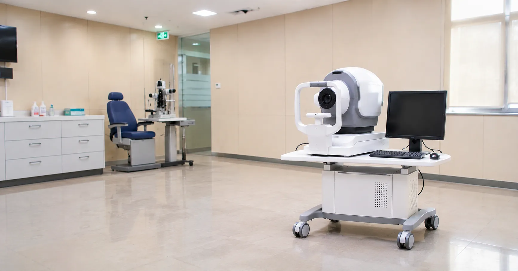
Optical coherence tomography (OCT) produces cross-sectional images of living tissue at near-microscopic resolution using light rather than sound or radiation. In eye clinics it is the standard way to image the retinal layers, optic nerve head and cornea, revealing structures a fraction of a millimetre thick without touching the eye. For a buyer the choice comes down to the imaging engine, the scan and analysis suite, throughput, connectivity and the total cost of running the platform.
The core answer is simple: OCT works like ultrasound but with light. It measures the echo time delay of reflected light to build a depth profile, and by scanning across the tissue it assembles those profiles into a detailed two- or three-dimensional image.
The measurement principle in detail
OCT relies on low-coherence interferometry. A beam of near-infrared light is split into a sample arm, which enters the eye, and a reference arm of known path length. Light reflected from different depths in the tissue recombines with the reference light, and interference occurs only where the two path lengths match. By analysing that interference the system determines how strongly each depth reflects, producing an axial scan — a single depth profile. Sweeping the beam across the retina stacks many axial scans into a cross-sectional B-scan, and repeated B-scans build a volume. Because the eye is transparent, near-infrared light penetrates to the retina and back, giving axial resolution of only a few micrometres, fine enough to separate the individual retinal layers.
Time-domain, spectral-domain and swept-source
Time-domain OCT
The original design mechanically moved the reference mirror to sample each depth in turn. It works but is slow, and it has largely been superseded in new purchases.
Spectral-domain OCT
Spectral-domain (Fourier-domain) systems use a fixed reference and a spectrometer to capture all depths at once, giving tens of thousands of axial scans per second and excellent retinal detail. This is the workhorse of most eye clinics.
Swept-source OCT
Swept-source systems use a rapidly tunable laser at a longer wavelength, scanning faster still and penetrating deeper through the retina to the choroid. They suit high-throughput clinics and deeper imaging, at a higher price. Decide which engine matches your caseload before comparing brands, because it drives both capability and cost.
OCT angiography and added modules
Many platforms add OCT angiography, which detects blood flow by comparing repeated scans of the same spot, mapping the retinal and choroidal vasculature without an injected dye. Anterior-segment modules image the cornea, angle and anterior chamber, and some systems combine OCT with a fundus camera or scanning laser ophthalmoscope. List the modules your clinicians will actually use, because each adds cost and training and an unused module is wasted capital.
Key specifications a buyer must check
Compare axial and transverse resolution, scan speed in axial scans per second, and the scan patterns and field of view on offer. Check the segmentation and analysis software: layer thickness maps, optic-nerve and ganglion-cell analysis, normative reference databases and, crucially, reliable change-analysis for monitoring progression over time. Confirm eye-tracking and image-registration quality, since these keep serial scans aligned and are central to detecting real change. Review the display, capture ergonomics for patients with poor fixation, and how well the system copes with media opacities such as cataract.
Standards, regulation and data protection
An OCT scanner is a medical device that must carry valid UKCA or CE marking with instructions for use. It is a light-emitting ophthalmic instrument, so laser and light-source safety follows the IEC 60825 and ophthalmic-instrument standards, general electrical safety follows IEC 60601-1 and electromagnetic compatibility IEC 60601-1-2. Because it stores identifiable images, data handling must meet UK GDPR and your information-governance rules. Ask for the declaration of conformity and confirm Great Britain registration. The MHRA regulates devices in the UK, NICE guidance informs how imaging supports eye-care pathways, and wider guidance sits on GOV.UK.
Calibration, maintenance and image quality
OCT platforms are largely self-calibrating, running internal checks on the reference arm and detector at start-up, but image quality still depends on a clean optical path. Routine care means keeping the objective lens and any headrest optics clean, following the manufacturer's recommended checks, and monitoring signal-strength indices so faint scans are re-taken rather than reported. Annual preventive maintenance by the manufacturer covers the light source, alignment and software updates. Keep the normative database and analysis software current, because progression analysis depends on consistent versions.
Decontamination and single-use considerations
Standard retinal OCT is non-contact, so between patients the chin rest and forehead band are wiped with an approved disinfectant, and disposable tissue or single-use covers may be used on the chin rest. Anterior-segment or intraoperative variants that touch the eye need a validated cleaning or a single-use cap per the instructions for use. Confirm which surfaces contact the patient and agree the wipe-down routine with infection control, keeping approved wipes at each station so throughput is not interrupted.
Care settings and connectivity
OCT is found in hospital ophthalmology and glaucoma clinics, high-street optometry practices, diabetic-eye-screening services and increasingly in community diagnostic hubs that refer into hospital eye services. Portability varies: most units are bench-mounted, though handheld OCT exists for theatre, bedside and paediatric use. Connectivity is decisive in a shared-care pathway — confirm DICOM output, integration with your ophthalmology image-management system and electronic patient record, and secure image sharing between community and hospital sites so a scan taken in the community can be reviewed by a hospital consultant without repeat imaging.
Consumables, total cost of ownership and servicing
OCT has modest consumables — cleaning wipes, chin-rest tissues and occasional printer supplies — but real lifetime costs sit in software licences, normative-database and module upgrades, annual service contracts and staff training. Ask suppliers for warranty length, whether the first year of service and software updates are included, repair turnaround, loan-unit availability and the cost of major parts such as the light source or spectrometer. A scanner out of action idles a clinic, so a dependable service route matters. Standardising platforms across sites simplifies training, comparison of serial scans and support. Our team can help you weigh the options — register as a buyer or contact us to compare specifications and availability.
Procurement checklist
-
Decide the imaging engine — spectral-domain or swept-source — for your caseload and depth needs.
-
Confirm the modules you need, such as OCT angiography or anterior-segment imaging, and drop those you do not.
-
Check scan speed, resolution, eye-tracking and the reliability of change-analysis for monitoring.
-
Verify normative databases and that segmentation software suits your clinical pathways.
-
Confirm UKCA/CE marking, IEC 60601 and light-safety compliance and certificates.
-
Check DICOM output and integration with your image-management system and patient record.
-
Agree the non-contact cleaning routine and any single-use covers with infection control.
-
Get warranty, software-upgrade and major-part costs and repair turnaround in writing.
Conclusion
OCT gives clinics a fast, non-contact window into the microscopic structure of the eye, and buying well means matching the imaging engine, modules and analysis software to the pathways you run. Weigh capability against lifetime software and service costs, confirm the regulatory and data-protection paperwork, and check that scans move cleanly between your systems and sites. Comparing verified specifications through MediGear helps your eye service invest in a platform clinicians will rely on for years.
Disclaimer
This article is for informational purposes only. It is published by MediGear (medigear.uk) for general information and procurement guidance, and is not clinical, diagnostic, treatment, technical, engineering, legal or regulatory advice, nor a product endorsement, guarantee or substitute for professional assessment. MediGear does not provide medical consultations. Buyers should consult their clinical, biomedical, estates and regulatory contacts, and the manufacturer's documentation, and independently verify all specifications, certifications, compatibility and suitability before purchase. Specifications, certifications and availability are correct at the time of publication and may change without notice. MediGear is a medical-equipment distributor and does not sell medicines or pharmaceutical products.