A duplex ultrasound system combines a real-time greyscale image with colour and spectral Doppler on the same screen, so a sonographer can see a vein and measure the blood moving through it at once. For venous assessment, that pairing is the whole point: the B-mode picture shows the vessel wall and any clot, and the Doppler shows whether blood is flowing, in which direction,n and how fast.
For a buyer, a duplex ultrasound system is a substantial capital purchase, and the venous work it will do — deep vein thrombosis scanning, checking patency, mapping the leg veins — sets what transducers and Doppler capability you actually need. This article stays on the general venous duplex study; reflux testing for varicose-vein surgery is a related but distinct assessment covered separately.
Duplex, triplex and how the modes combine
Duplex means two modes together: B-mode anatomy and Doppler flow. Add a colour overlay, and many call it triplex, though the industry still says duplex. The greyscale image lets the operator find a vein and, crucially, compress it — a normal vein flattens under gentle probe pressure, a thrombosed one does not, and that compression test is the backbone of a deep vein thrombosis scan. Colour Doppler paints flow onto the image so absent or abnormal flow is obvious at a glance, and spectral Doppler at a sample gate gives the velocity trace and the respiratory phasicity that confirms a vein is patent above the point being scanned.
Transducers that suit venous work
Probe choice defines the study. A linear-array transducer, broadly in the 5–12 MHz range, is the mainstay for leg veins, giving the high-resolution near-field image needed to see the vein wall and compress it. For deep pelvic or iliac veins, or a large oedematous limb, a lower-frequency curvilinear probe penetrates further at the cost of resolution. That penetration-versus-resolution trade-off is the core transducer decision in venous work: high frequencies resolve a superficial vein wall crisply but fade with depth, while a 2–5 MHz curvilinear probe reaches the iliac and pelvic veins at the expense of fine detail. A venous service therefore needs at least a good linear probe and often a curvilinear one. Confirm which transducers are included, the number of active probe ports, and that the frequencies match the depth of the veins you scan.
The Doppler settings that make a venous study work
Venous flow is slow, so the machine must be tuned for it. A low pulse-repetition frequency and a low wall filter let the system show sluggish venous flow that arterial presets would strip out, while the colour scale, gain and Doppler angle all have to be set for low velocities. Augmentation is central: a distal calf or thigh squeeze should send a burst of flow up a patent vein, respiratory phasicity confirms an unobstructed segment above, and a Valsalva manoeuvre loads the sapheno-femoral junction. Separate, well-labelled presets for a two- or three-point DVT compression study and for full saphenous mapping save reconfiguring the machine mid-list. Test that venous workflow, not just the arterial one, when you trial a machine.
Cart, portable and point-of-care formats
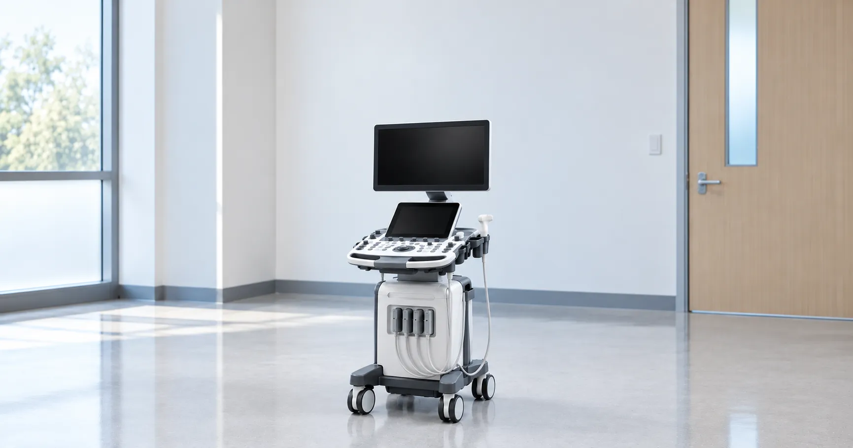
Duplex ultrasound systems range from full cart-based consoles to laptop-style portables and compact point-of-care units. A vascular lab running full mapping and complex studies wants a cart with the best image processing and the widest probe support. A ward-based or emergency deep vein thrombosis service may be well served by a portable duplex system that reaches the bedside. Point-of-care units suit focused two-point or three-point compression scans but are not a substitute for a full diagnostic study. Match the format to where and how detailed your venous scanning is.
What venous duplex is used to answer
The venous duplex study answers a defined set of questions. Is there a deep vein thrombosis, and if so, where,e and is it occlusive? Is a vein patent and compressible along its length? What is the venous anatomy before an intervention or an access procedure? Vascular labs, emergency departments, and DVT clinics all lean on it because it is non-invasive, repeatable, and radiation-free. NHS pathways route suspected clot to duplex scanning, and referrers can read the care context at nhs.uk; the buyer's job is to specify a system that answers these questions cleanly at the point the scan is done.
Standards, marking and electrical safety
As diagnostic ultrasound equipment, a duplex ultrasound system is built to the general standard IEC 60601-1, with the particular standard for ultrasonic diagnostic equipment, IEC 60601-2-37, governing acoustic output and safety. It must carry UKCA or CE marking, and the supplier should provide conformance documentation you can file. Regulation of the duplex system and any field-safety notices rests with the Medicines and Healthcare products Regulatory Agency at mhra.gov.uk. This guide draws on manufacturer specifications, the IEC 60601 family and UK regulatory guidance, and is written for UK procurement and clinical-engineering teams.
Probe hygiene and everyday care
Linear and curvilinear probes touch intact skin, so they need cleaning and low-level disinfection between patients using only the manufacturer's approved agents; the wrong chemical cracks the probe lens and voids the warranty. Coupling gel is a steady consumable, and single-use sachets help where cross-contamination is a concern. Confirm the compatible-disinfectant list before standardising your wipes, and build a documented cleaning protocol around the machine, because probe damage from incorrect cleaning is one of the commonest avoidable costs.
Connectivity, DICOM and reporting
A venous study is only useful once it reaches the record. Confirm the duplex ultrasound system exports DICOM cleanly to your PACS and vascular reporting system, supports structured reporting with the measurements a venous study needs, and handles worklists so images attach to the right patient automatically. Storage capacity, wired or wireless transfer, and the reporting package all shape how quickly a sonographer turns a list around. Integration gaps are a common and avoidable drag on a busy vascular service, so test the whole workflow, not just the image.
Image quality is where money is spent.t
A venous duplex study lives on image quality: a clear vein wall to judge compressibility, sensitive colour Doppler to catch slow flow, and clean spectral traces. Better transducers, more processing channels, and tissue harmonic imaging all sharpen the difficult study — the oedematous leg, the deep vein, the large patient — which is exactly where a basic machine fails. When you trial a system, scan a genuinely difficult limb rather than an easy volunteer, because the gap between an entry-level and a capable duplex system shows most on the hard cases that fill a real venous list.
Beyond the purchase price
For a venous duplex system, the base console is only a fraction of the eventual spend. Transducers are expensive, get dropped, and eventually fail, so weigh probe warranty and replacement terms heavily. Budget for the service contract, software upgrades, coupling gel, cleaning consumables, es and the eventual battery replacement on portable units. A well-supported refurbished cart from a reputable supplier can stretch a capital budget if it comes with warranty, current software and a service history. Compare whole-life cost, not headline price, and use MediGear to weigh suppliers on equal terms.
Training and the operator's role
Venous duplex is highly operator-dependent, so the machine is only half the investment. A reliable deep vein thrombosis scan depends on correct probe pressure for the compression test, patient positioning, and skilled use of augmentation and Doppler angle. A department adding or replacing a duplex ultrasound system should plan sonographer training, a protected scanning list, and agreed local protocols for which vein levels are assessed. A capable machine in untrained hands still produces unreliable studies, so budget the training alongside the hardware, not after it.
Specifying a venous duplex system: buyer's checklist
-
A high-frequency linear transducer for leg veins, plus a curvilinear probe for deep or oedematous limbs.
-
Colour and spectral Doppler tuned for slow venous flow, with quick, well-labelled venous presets.
-
Enough active probe ports and confirmation of which transducers are included in the price.
-
Format fit — cart for the vascular lab, portable for bedside DVT scanning.
-
Documented conformance to IEC 60601-2-37 for the duplex system, plus a current UKCA or CE mark you can verify.
-
Clean DICOM export to PACS and your vascular reporting system, with worklist support.
-
Transducer warranty terms and the true whole-life cost, taking in service cover, software upgrades and consumables.
The verdict on venous duplex
A duplex ultrasound system is the mainstay of venous assessment because it shows the vein and measures its flow together, answering the deep vein thrombosis, patency and mapping questions in one non-invasive study. Buy it around the transducers, the slow-flDoppler, and the reporting workflow, and remember it is only as good as the sonographer using it. If you are specifying a venous duplex system, MediGear can help you compare verified suppliers and probe options side by side.
Disclaimer
This article is for informational purposes only. It is published by MediGear (medigear.uk) for general information and procurement guidance, and is not clinical, diagnostic, treatment, technical, engineering, legal or regulatory advice, nor a product endorsement, guarantee or substitute for professional assessment. MediGear does not provide medical consultations. Buyers should consult their clinical, biomedical, estates and regulatory contacts, and the manufacturer's documentation, and independently verify all specifications, certifications, compatibility and suitability before purchase. Specifications, certifications and availability are correct at the time of publication and may change without notice. MediGear is a medical-equipment distributor and does not sell medicines or pharmaceutical products.