A craniotomy is the surgical removal of a section of skull, the bone flap, to reach the brain, after which the flap is replaced and fixed back in place at the end of the operation. It is the standard route for tumour removal, evacuating a bleed, clipping an aneurysm and many other neurosurgical procedures. The instruments fall into a clear sequence: opening the scalp, making the bone flap, opening the dura, working within the brain, and closing and refixing the bone.
Neurosurgical instruments are precise, expensive, and heavily reliant on powered systems and fine hand tools, so procurement here is as much about the drill platform, its consumables, and its servicing as about the trays. This guide walks through the instrument groups a neurotheatre needs and what a buyer should check when specifying them.
How a craniotomy is structured
After positioning and often rigid head fixation, the surgeon opens the scalp and reflects it, then makes the bone flap. Small holes, burr holes, are drilled through the skull, and a cutting tool joins them to free a flap of bone, which is lifted off to expose the dura. The dura is opened to reveal the brain, t;e. The cranial work is done under magnification, and the dura is closed, the bone flap is replaced and fixed, and the scalp is closed. Each stage has its own instruments, and the whole procedure depends on rigid fixation, powered bone tools and fine microsurgical instruments working together.
Head fixation and positioning
The head is usually held immobile in a skull clamp with sterile pins that grip the skull at fixed points, giving the stability micro-neurosurgery demands and a frame to which retractor systems and navigation can attach. Buyers should confirm the clamp, its single-use or reusable pins, and the adaptors that link it to the operating table and to any self-retaining retractor and navigation platform, since these must all interface correctly.
Powered bone tools: the craniotome and perforator
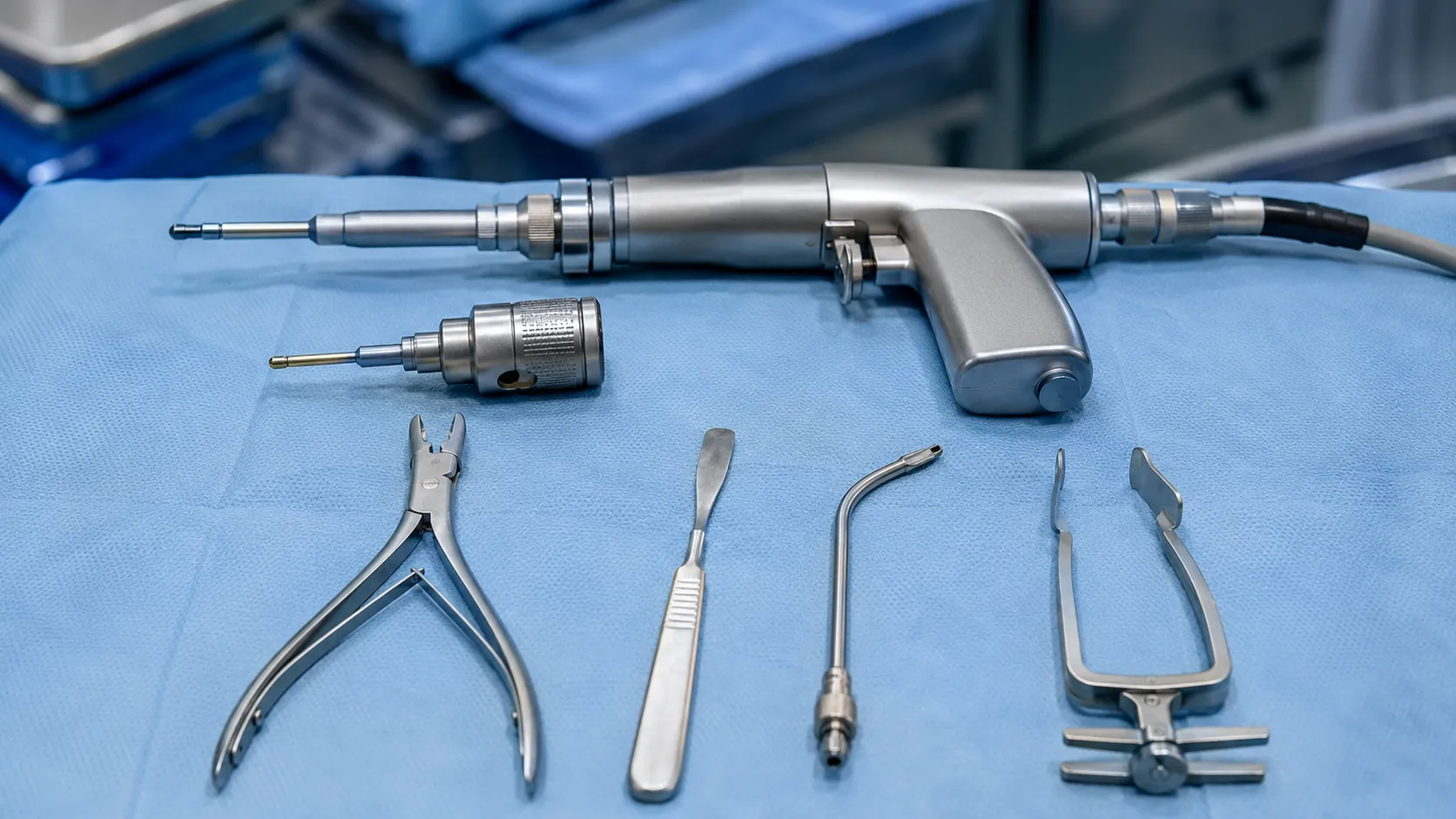
The heart of the set is the powered drill system. A pneumatic or electric high-speed drill runs a range of attachments: a perforator burr that drills the initial hole and is designed to stop before it plunges through into the brain, and a craniotome attachment with a footplate that slides under the bone to cut between holes and raise the flap while shielding the dura. Various burrs and cutting bits handle bone removal and shaping. Where power is not used, a manual Hudson brace and Gigli saw with guide can make burr holes and cut bone. Buyers must specify the drill platform, its handpieces and attachments, the single-use perforators and burrs, the irrigation that cools the bit, and the service and repair cover, because the drill is the item most likely to halt a list if it fails.
Opening the dura and controlling bleeding
Once the flap is off, fine tools open the dura: a dural hook and fine forceps lift it, and small scissors incise it. Haemostasis is constant and meticulous. Bipolar forceps, powered by an electrosurgical generator, coagulate tiny vessels precisely without spreading current, and are fundamental to neurosurgery; a range of tip sizes and non-stick options is usually held. Bone-edge bleeding is controlled with bone wax, and dural and cortical bleeding with haemostatic agents and patties. Buyers should stock a range of bipolar forceps, the generator that drives them, and the haemostatic consumables the surgeons prefer.
Microsurgical and retraction instruments
The intracranial work is performed using one-handed instruments under an operating microscope. Dissectors such as Penfield and Rhoton patterns separate tissue planes; micro-scissors, micro-forceps and fine suckers work in a small, deep field. Rongeurs (Kerrison and Leksell patterns) nibble away bone, and periosteal elevators strip tissue from bone. Self-retaining retractor systems, often mounted to the head clamp, hold the field open and can carry brain retractors. Aneurysm and specialist work adds clips and appliers. Buyers should ensure that microsurgical trays, the retractor system and its mountings, and special-d-ons are complete, standardised, and compatible with the head fixation in use.
Bone-flap fixation and closure
At the end, the bone flap is replaced and secured, historically with sutures but now usually with low-profile titanium plates and screws or dedicated cranial fixation clamps that hold the flap flush and stable. The dura is closed, sometimes with a graft or substitute, and the scalp is closed in layers. Fixation devices are implants, so buyers must confirm they are UKCA- or CE-marked, I-compatible where stated, and traceable by batch, and that the screwdrivers and appliers for the chosen system are in the tray.
Standards, regulation and decontamination
Reusable neurosurgical instruments must be decontaminated through a validated process between cases, and neurosurgery attracts particular attention to instrument tracking and decontamination because of the theoretical risk associated with central nervous system tissue; single-use is preferred for some items on that basis. Powered tools and electrosurgical generators are active electrical equipment governed by the IEC 60601 family. Cranial fixation plates, single-use perforators, and burrs are medical devices that require A or CE marking; implants must never be reprocessed. The MHRA regulates all of these and issues safety guidance, with clinical standards available from bodies such as NICE. Maintain full traceability for instruments and implants.
Consumables, servicing and total cost of ownership
Neurosurgical capital, the drill system, microscope interface, head fixation and retractor platform, is a major investment, and its total cost of ownership is dominated by service contracts, handpiece maintenance and spares. The recurring consumable costs are use-perforators, burrs, patties, haemostatic agents and fixation implants. A robust service and repair arrangement for the drill and generator is essential, since downtime cancels complex lists. Standardising fixation systems and bipolar patterns across theatres simplifies stock and training. We can help you specify a neurosurgical set and its service cover — register as a buyer or contact our team to discuss requirements.
Use across care settings.
Craniotomy is concentrated in neurosurgical centres and major trauma units, not hospitals; hospitals' equipment is located in high-dependency theatres. A tertiary neuroscience centre runs several neurotheatres and needs redundancy in drills and generators so a single failure never stops emergency work, plus deep stock of consumables and fixation systems. A trauma unit performing emergency evacuation of bleeds needs a reliable core set and immediate availability out of hours. Matching capital redundancy and consumable depth to caseload keeps neurosurgery running safely.
Procurement checklist
-
Specify the powered drill platform with perforator and craniotome attachments, plus service cover.
-
Confirm single-use perforators, burrs and irrigation are stocked and compatible with the handpieces.
-
Check the head clamp, pins and adapters, tors adaptor interfaces,s retractors, and navigation.
-
Hold a range of bipolar forceps and the generator that drives them, plus haemostatic consumables.
-
Ensure microsurgical trays, dissectors, rongeurs and the retractor system are complete and standardised.
-
Confirm cranial fixation plates are UKCA or CE marked, MRI-compatible where stated, and batch-traceable.
-
Plan drill and generator redundancy so a single failure never cancels emergency lists.
Conclusion
A craniotomy set is best understood as a sequence: fixation and positioning powered bone tools to raise the flap; fine instruments and bipolar forceps to open the dura and control bleeding; microsurgical tools for the cranial work; and fixation plates to refix the bone. The buying decisions that matter most are the drill platform and its service cover, the bipolar and generator pairing, and the compatibility of head fixation, retractors and fixation implants. MediGear can help specify and supply the neurosurgical instruments and consumables a brain-access service depends on, with the servicing that keeps them ready.
Disclaimer
This article is for informational purposes only. It is published by MediGear (medigear.uk) for general information and procurement guidance. It is not clinical, diagnostic, treatment, technical, engineering, legal or regulatory advice, nor a product endorsement, guarantee or substitute for professional assessment. MediGear does not provide medical consultations. Buyers should consult their clinical, biomedical, estates and regulatory contacts, and the manufacturer's documentation, and independently verify all specifications, certifications, compatibility and suitability before purchase. Specifications, certifications and availability are correct at the time of publication and may change without notice. MediGear is a medical-equipment distributor and does not sell medicines or pharmaceutical products.