
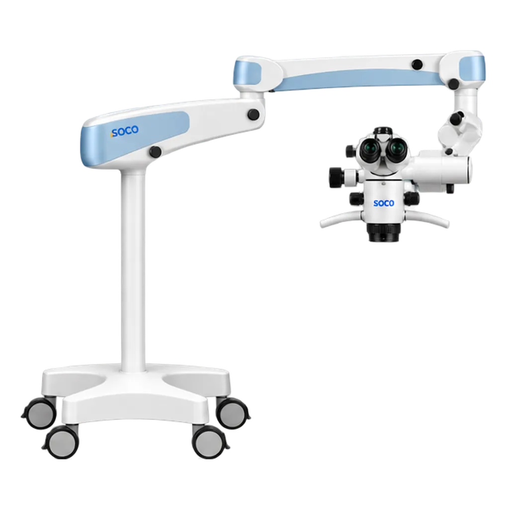
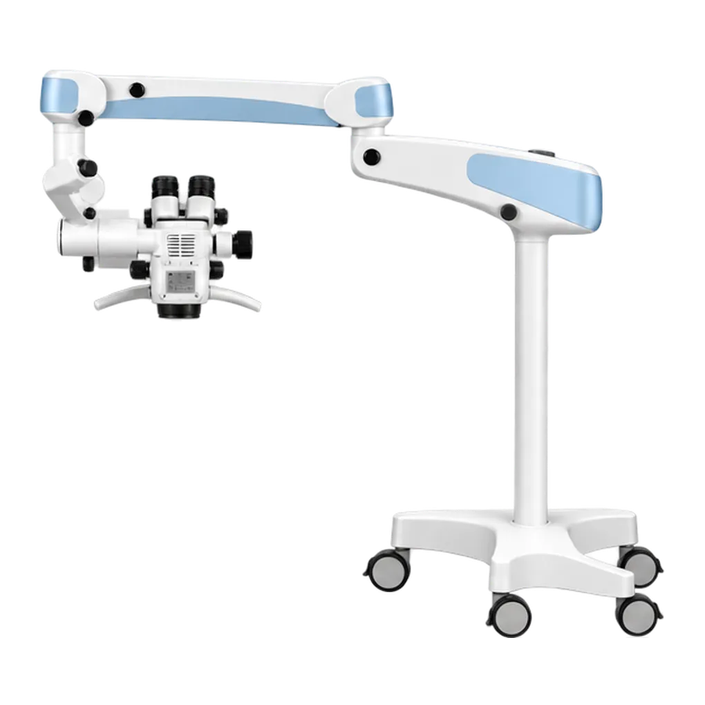
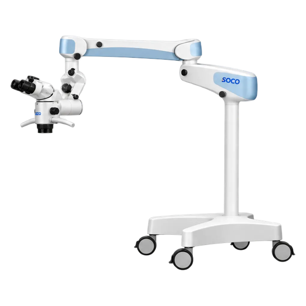
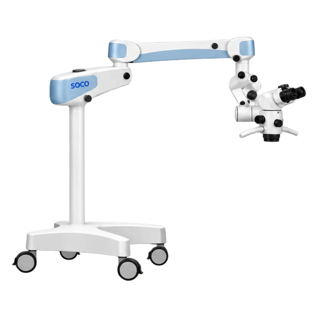
The SCM800-UL suits practices where clinical photography is part of everyday work. Endodontists photograph canal anatomy, separated instruments, perforation repairs and obturation results for records and referral reports. Restorative and aesthetic dentists capture preparation margins, shade matching and finished restorations in high-resolution stills. A DSLR's sensor and colour handling make those images suitable for portfolios, publications and lectures.
Chairside communication is the second major use. With live images sent over HDMI to a monitor, the clinician shows the patient a cracked tooth, a carious lesion or a completed restoration as it appears under magnification. Patients see the detail behind the treatment plan, which supports informed consent and increases treatment satisfaction.
Teaching and peer review make up the third. Recorded procedures show technique exactly as the operator saw it, from the operator's own viewpoint at the microscope. Dental schools, postgraduate courses and study clubs use live HDMI output to show procedures to a room or to remote viewers. The recorded material supports academic exchange and case presentations. Periodontal and implant surgeons record microsurgical techniques for training, using the green filter to show vessels and nerves clearly on camera.
Its users include endodontists, aesthetic and restorative dentists, practices with a strong referral network, clinical educators and dentists who publish or lecture. Clinicians who already own a Canon or Sony camera gain the most, because the standard interface lets them use it on the microscope straight away. The 195 to 450 mm working distance, 0° to 180° binocular tube and pendulum system keep the clinician comfortable through long recorded sessions.