
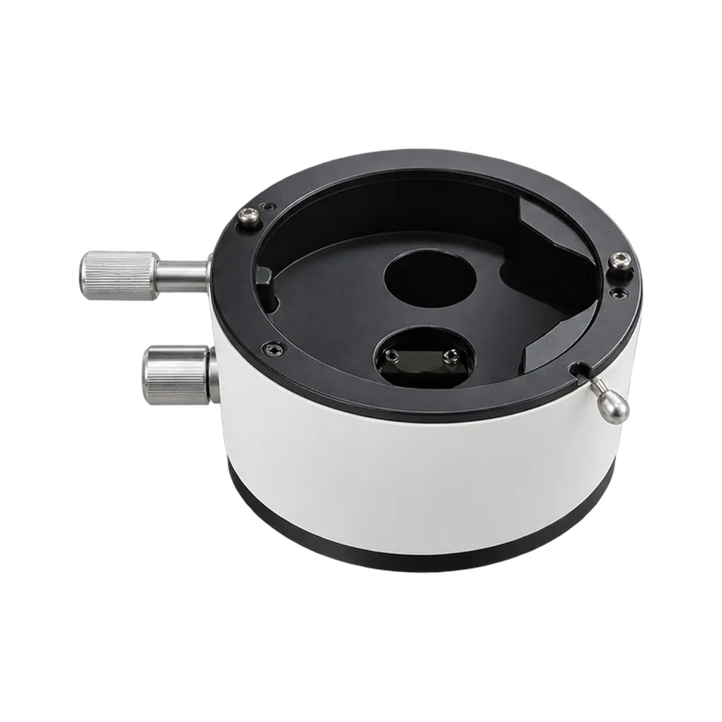
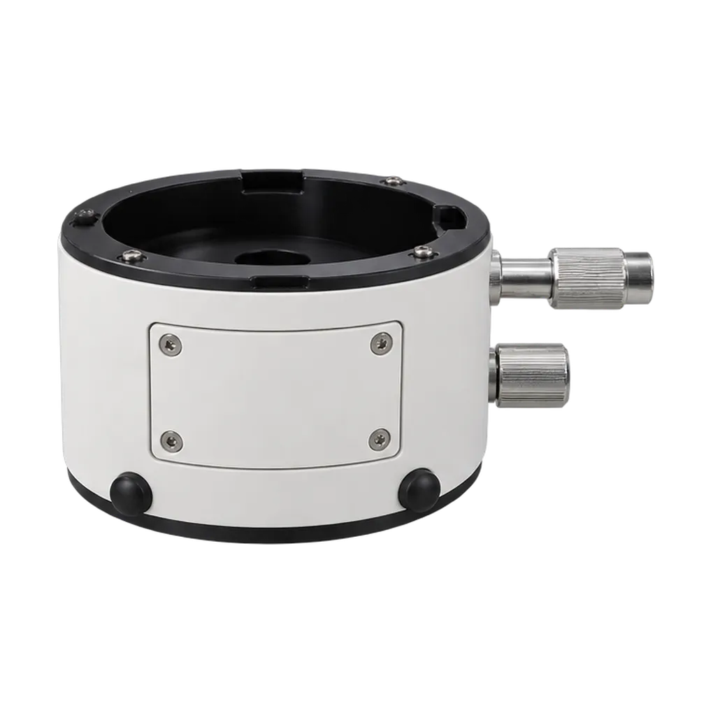
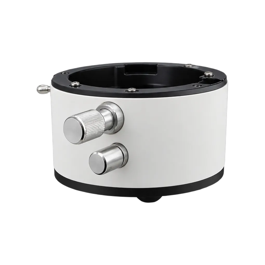
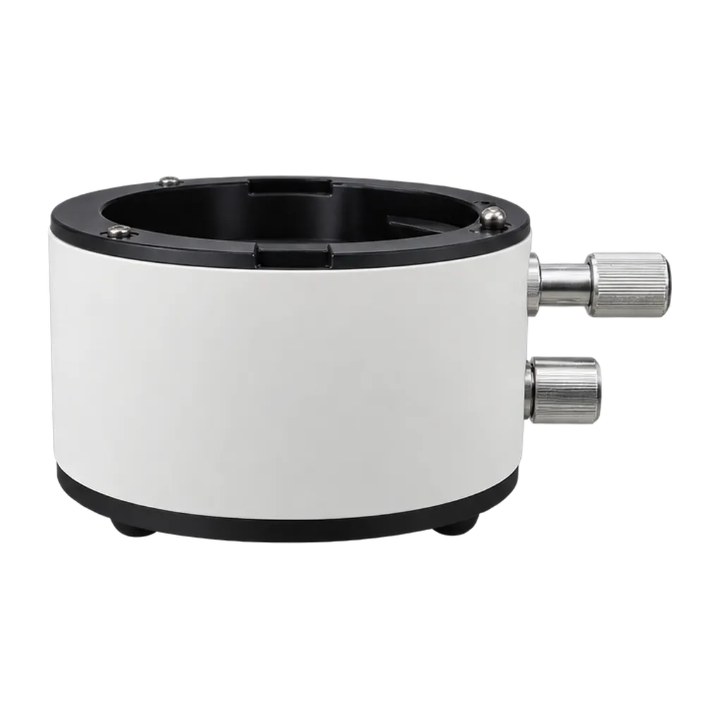
The BZ1.6 belongs to the stages of treatment where the microscope's normal top magnification is just short of what the eye needs. Endodontics provides the clearest examples. Locating a second mesiobuccal canal in an upper molar, finding a calcified orifice or tracing the isthmus between two canals all reward the extra step. The clinician engages the magnifier for those few minutes, then returns to the normal range for instrumentation and irrigation.
Crack detection is another strong use. A hairline fracture running across the pulp floor or down a canal wall can be hard to confirm at standard settings. The added 1.6× rate, combined with the microscope's light and filters, helps the clinician follow its full course. Restorative dentists use the same step to inspect the finish of a margin, the fit of an inlay or the seal of a bonded restoration.
In periodontal and implant microsurgery, the magnifier supports the finest suturing. Placing small sutures in papillae or closing a flap around an implant needs precise needle placement, and an extra step of enlargement helps with each pass. Teaching clinics can also use the added detail when demonstrating fine anatomy through the microscope's imaging system, giving students a clearer view of structures they will later need to find themselves.
Its users include endodontists, restorative specialists, periodontists, implant surgeons and educators using SOCO's SCM860, SCM850 Pro, SCM800 or SCM600 microscopes. Practices that already own one of these models can add the magnifier as their caseload moves towards more complex endodontic and microsurgical work, extending the working range of the instrument they already rely on.