
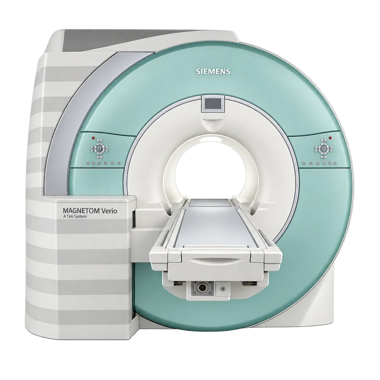
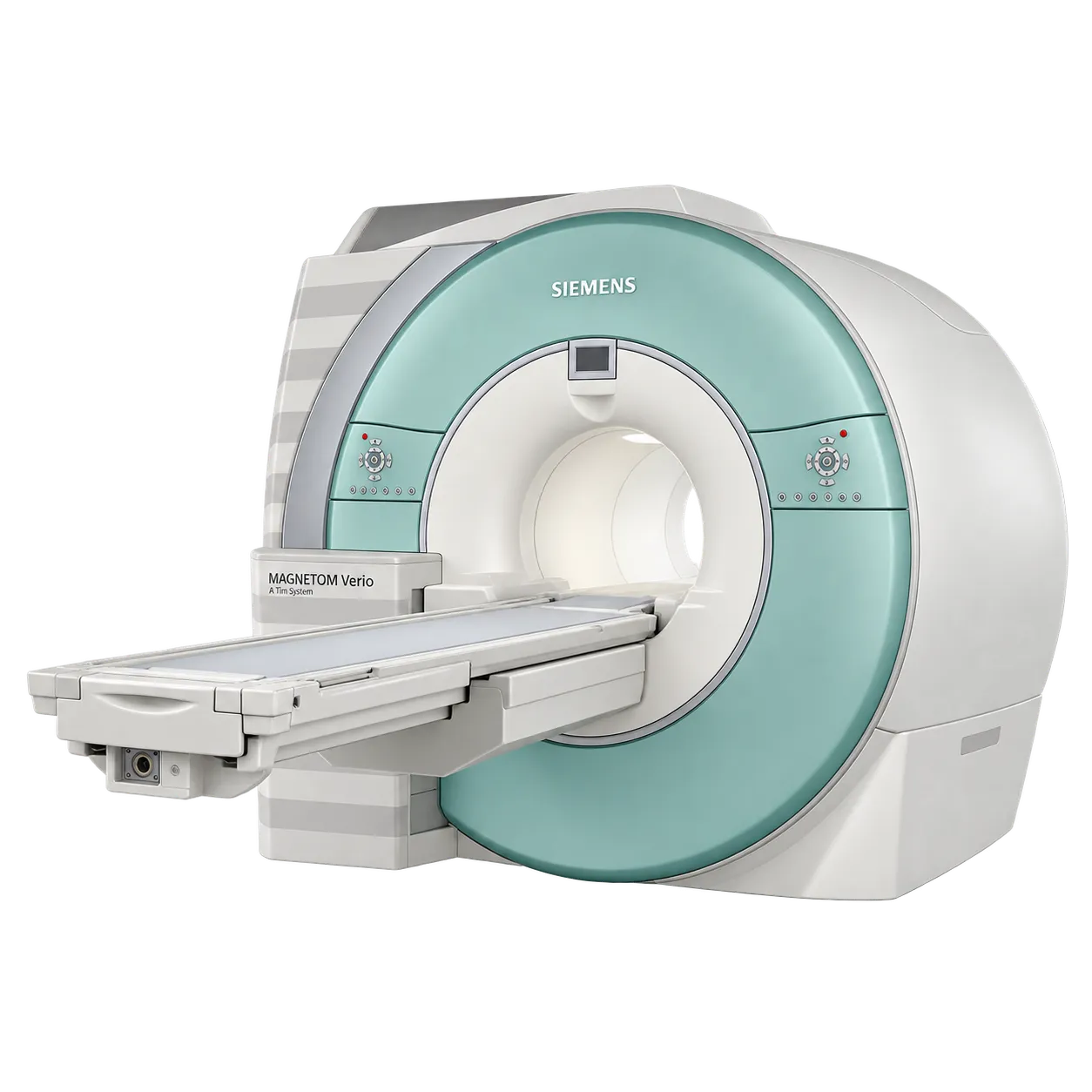
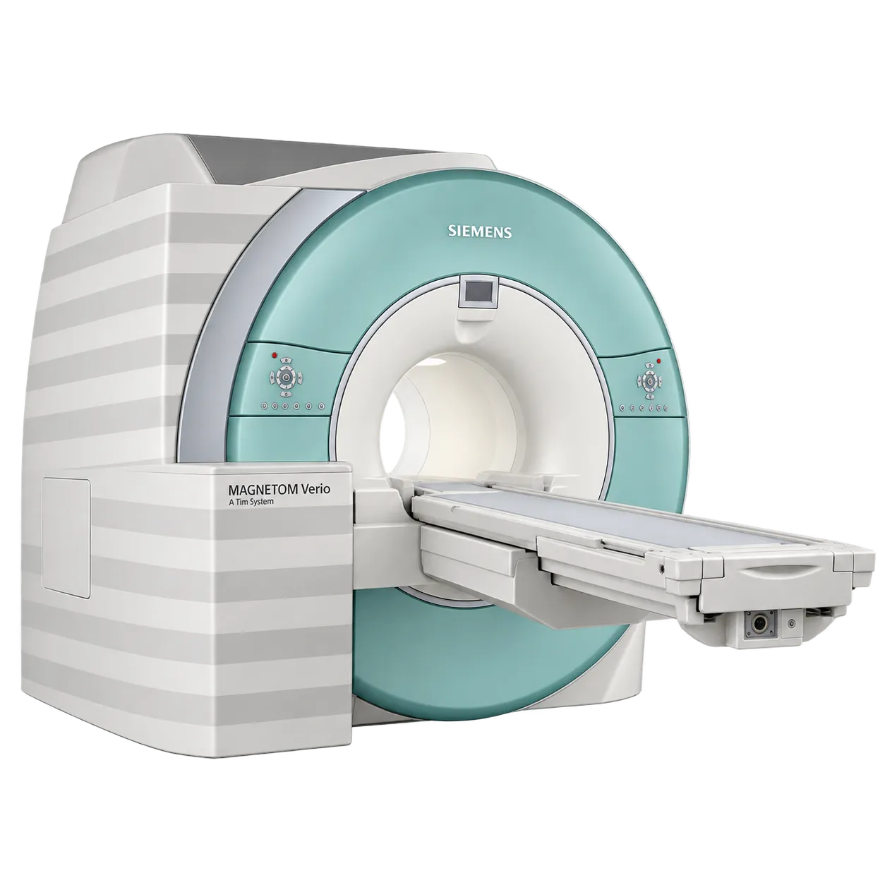
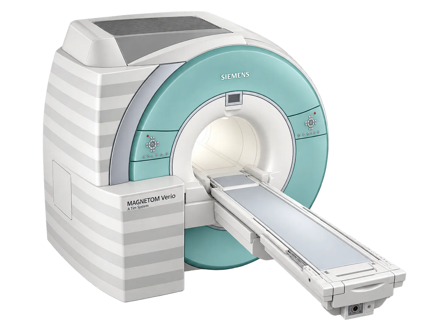
Neurological imaging is supported by head and neck coil elements combined into the imaging matrix, with parallel acquisition shortening the sequences a routine brain study relies on.
Spine imaging draws on integrated matrix elements along the table, with coverage extending without repositioning between regions.
Body and abdominal imaging runs within the 45 by 45 by 30 cm homogeneity volume, with patient-specific shimming applied per study.
Musculoskeletal imaging uses dedicated joint and extremity coils according to the set supplied with this system.
Angiographic and head-to-toe work is supported by parallel acquisition in all directions with PAT factors to eight.
Sites with restricted floor loading or limited plant capacity are served by a 4.3 tonne system with contained cooling and power requirements.
Services fitting an MRI into an existing building are helped by a 30 square metre minimum room area, a 2.4 metre ceiling and a fringe field held to 2.5 metres laterally.