
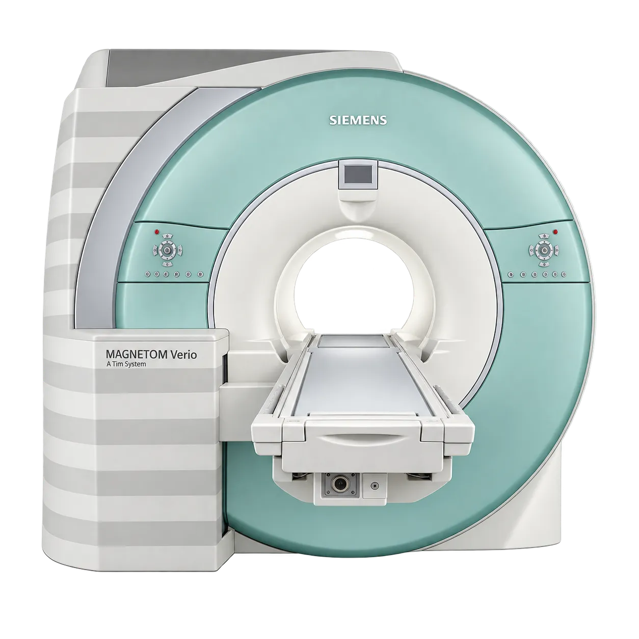
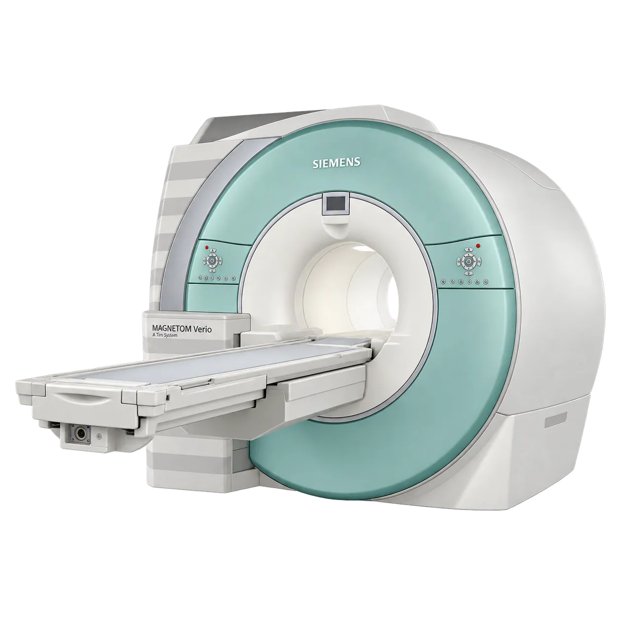
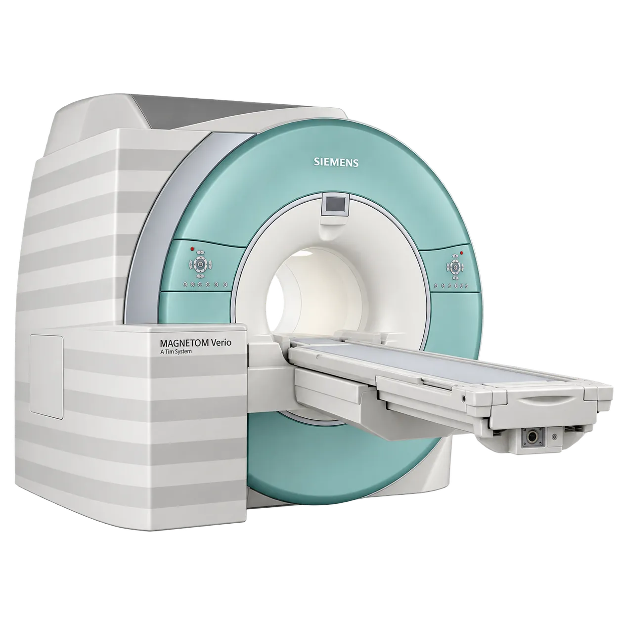
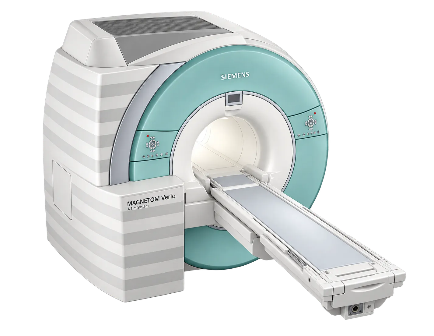
Neurological imaging benefits directly from guided workflow, since brain protocols are performed repeatedly and small differences in slice geometry between operators affect comparison at reporting.
Follow-up and surveillance studies are supported by the same guidance, because an examination acquired to a stored definition can be matched against the study before it.
Spine imaging draws on integrated matrix coil elements with coverage extending between regions without repositioning the patient.
Body and abdominal imaging is supported by strategies within the workflow engines that cover breath-hold variation, alongside patient-specific shimming applied per study.
Musculoskeletal imaging uses dedicated joint and extremity coils according to the set supplied with this system.
Angiographic and head-to-toe work runs on parallel acquisition in all directions with PAT factors to eight.
Departments training or rotating staff are supported by guidance that carries a defined method through the examination, so experience level has less bearing on the dataset produced.