
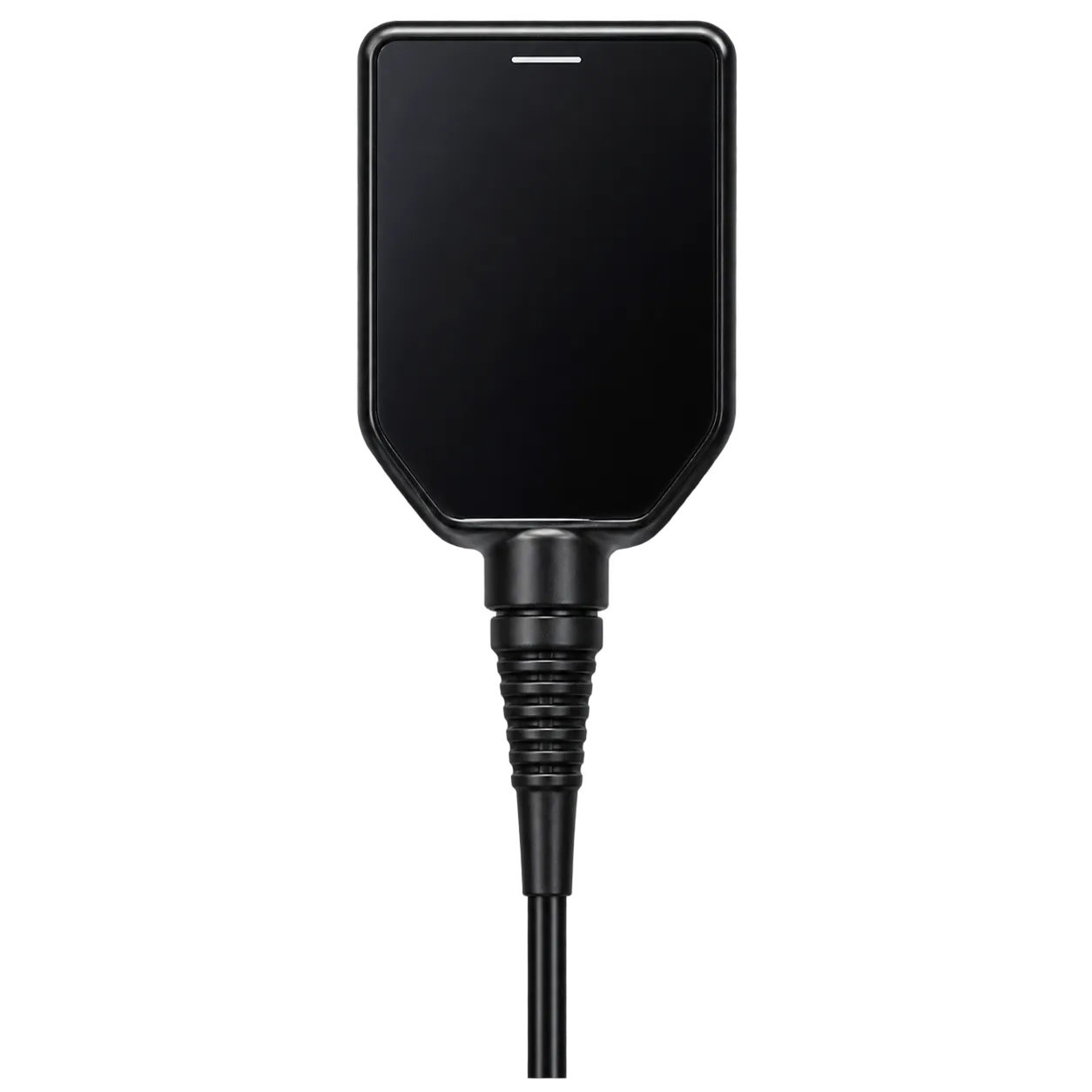
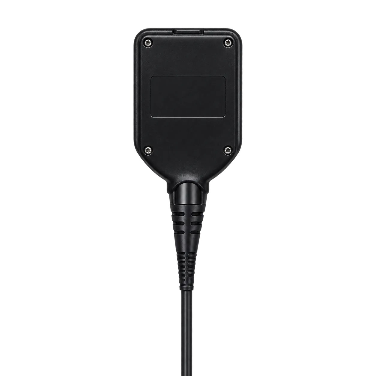
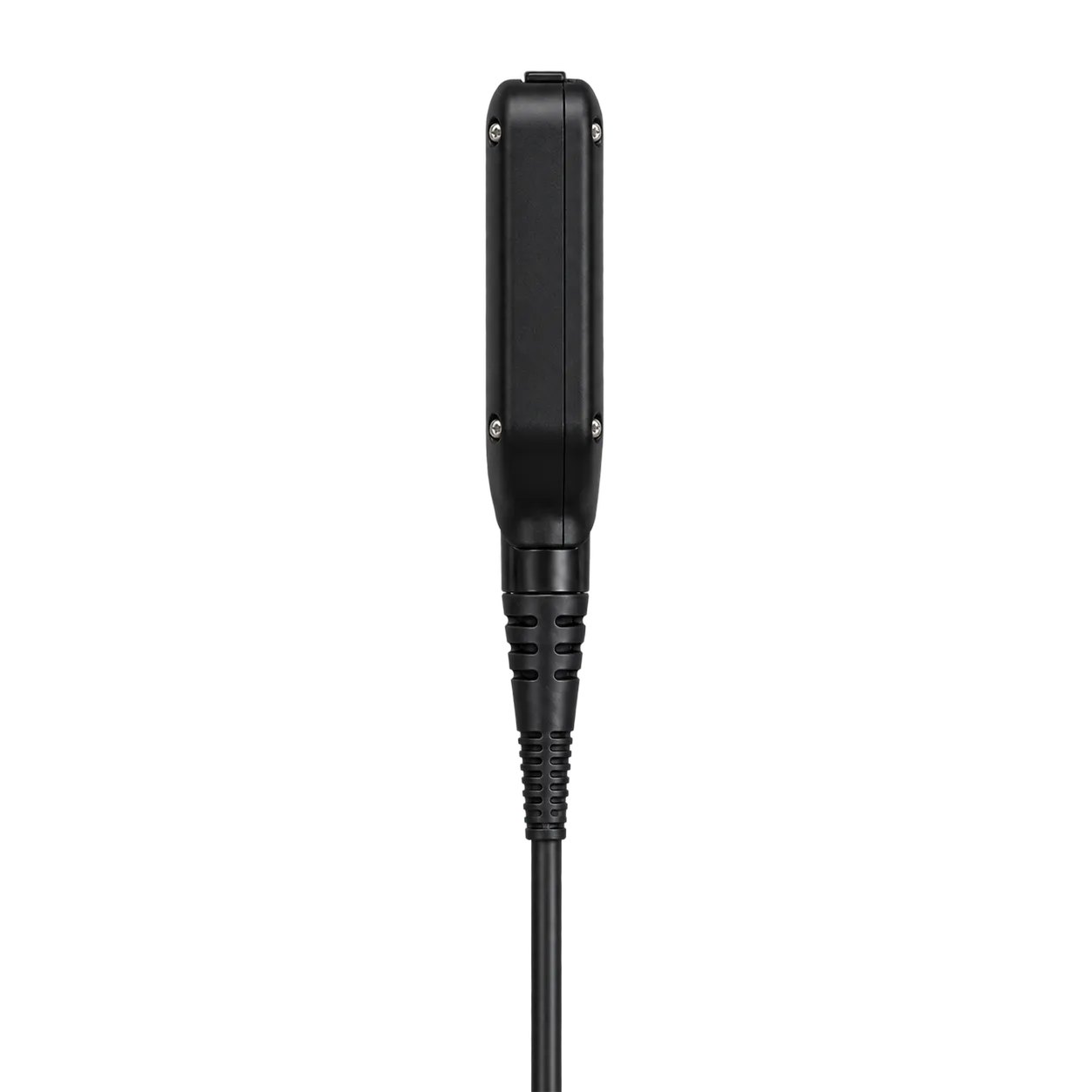
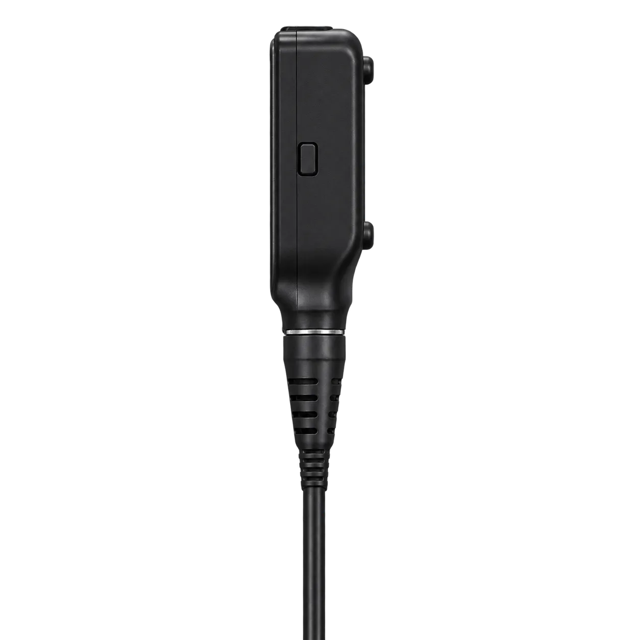
The URF-V serves urology departments, endoscopy suites and operating theatres where the ureter and kidney are examined and treated through a flexible route. Urologists and trained medical personnel operate it, and the work covers diagnosis, inspection of the collecting system, and treatment, with accessories passed along the 3.6 Fr channel and laser work supported by fine tip control.
A procedure begins with the 8.5 Fr distal end entering the ureter, and the 9.9 Fr tube follows as far as the 670 mm working length allows. The CCD chip at the tip forms the picture; the integrated cable carries it to the VISERA ELITE OTV-S190 processor, and the team watches it on a monitor. Steering then does the main work. The tip bends 180° up and 275° down, so the operator turns it to face each part of the collecting system in turn. The 90° forward view stays sharp from 2 to 50 mm, which suits both close inspection of tissue and a look across a broad space. When a laser fibre is in use, the insertion tube rotates 90° to either side to finely adjust the tip position. Narrow band imaging is available with a compatible light source, so the same scope serves both the standard view and the NBI view.
Four programmable buttons keep the picture under the operator's hand throughout. Image capture, white balance and zoom can be assigned to them, so a frame is saved or the view is adjusted without leaving the control section. Between cases, the scope supports leakage testing, and the integrated camera and light guide cable stay with it as one piece.