

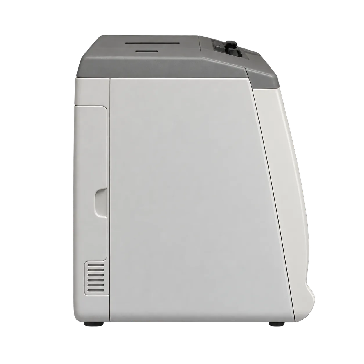
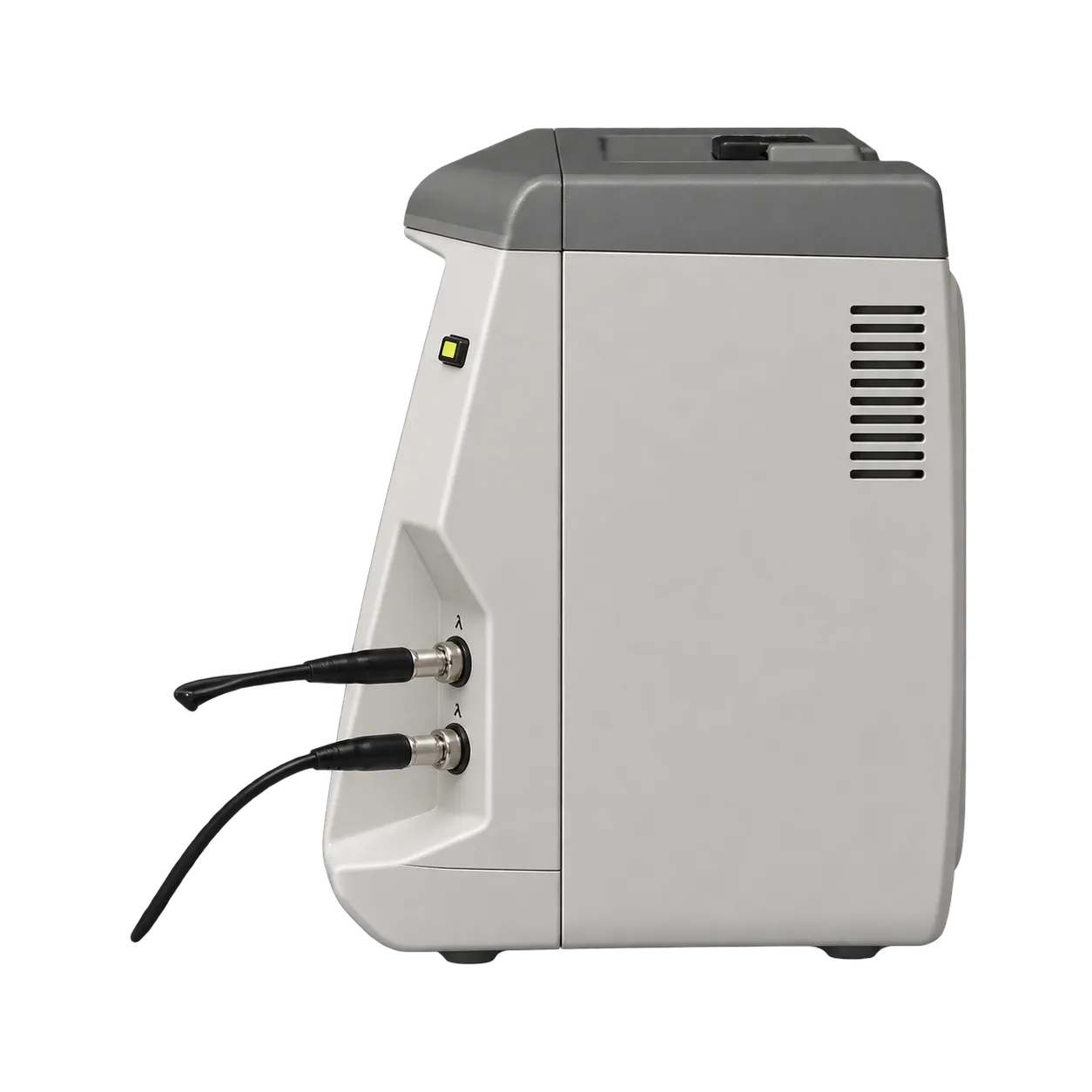
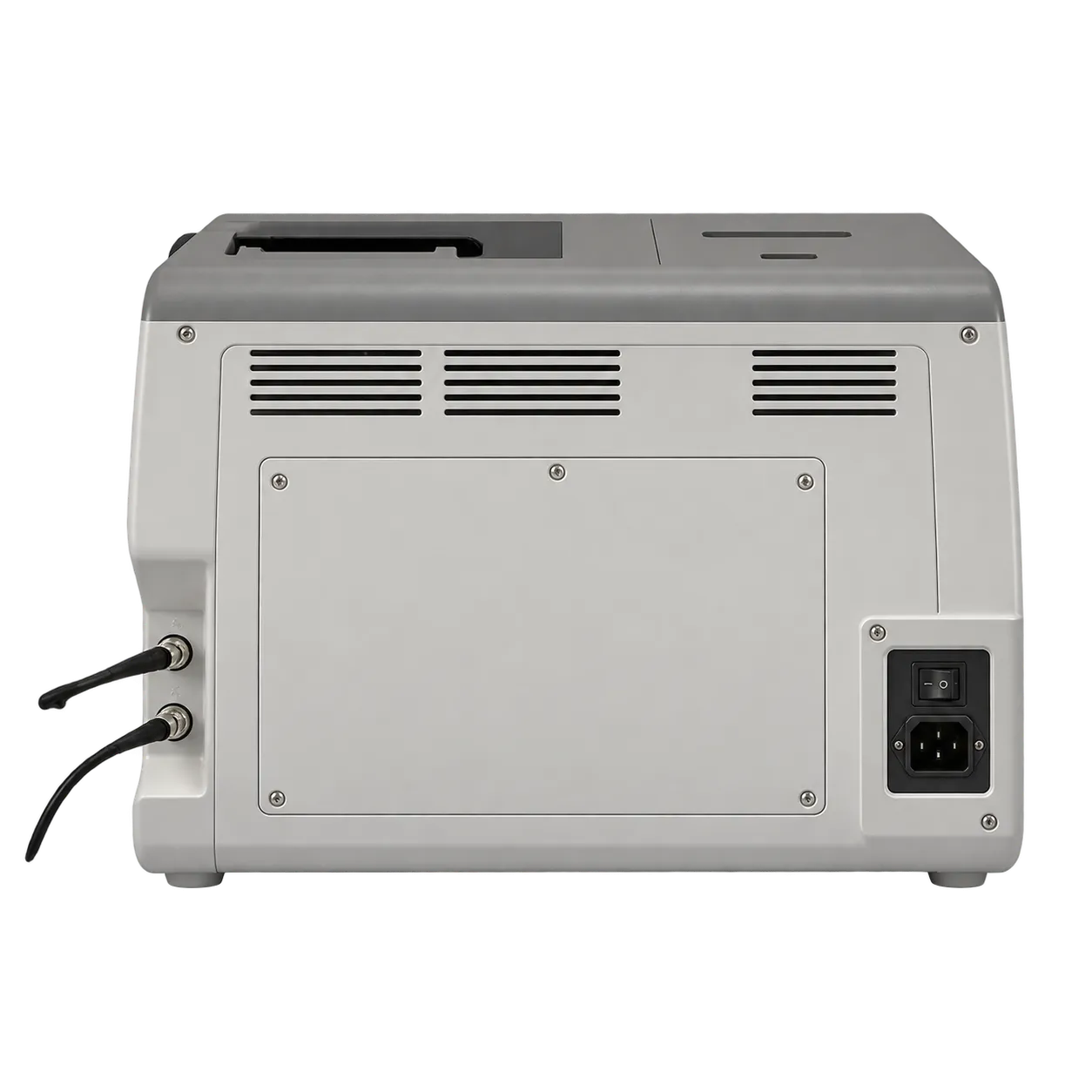
Cataract assessment is the principal setting for the US-1800's axial measurement and IOL calculation functions. Ophthalmologists and trained ophthalmic staff acquire the ultrasound trace, assess its tissue echoes, and select the reading used for implant planning. The instrument carries that result into a calculation with corneal curvature, IOL constants, and the intended postoperative refraction. Foot control supports this sequence by leaving the operator's hands available for positioning during capture.
Phakic, aphakic and implanted eyes have dedicated measurement settings. In an eye containing its natural lens, the displayed distances describe the anterior chamber, lens,s and vitreous segment as well as overall axial length. The selected mode supplies the acoustic conversion framework for the examination. Its relevance lies in matching the calculation to the ocular condition being measured, with the waveform available for professional assessment.
Corneal and refractive assessment uses the optional pachymetry function. Point-by-point thickness measurements support examinations before and after corneal refractive procedures and the assessment of corneal swelling. An editable location map relates the reading to its acquisition position. Repeated samples at each site produce a mean and standard deviation, giving the clinician both a representative thickness and a description of variation within that local series.
Within an ophthalmic department, saved traces, numerical lists and printed calculations support subsequent review of the same examination. Patient identifiers and the saved date organise the retained records. The compact tabletop console houses acquisition controls, calculation tools and printing together, while the reference test piece forms part of preparation before use. This combination suits a supervised measurement station where capture, interpretation and documentation remain closely connected.