
Neurological imaging runs on head and neurovascular phased array coils, with waveform repeatability bearing directly on diffusion-weighted sequences that acquire across many repetitions.
Spine imaging draws on high-density array coils placed along the region of interest, with the receive chain accommodating the coil supplied.
Body and abdominal imaging is supported across the 8-channel receive chain with array coils covering the region under examination.
Musculoskeletal imaging uses dedicated joint and extremity phased array coils according to the set supplied with this system.
Angiographic work depends on gradient timing accuracy, since flow encoding is defined by the shape and timing of the applied gradients.
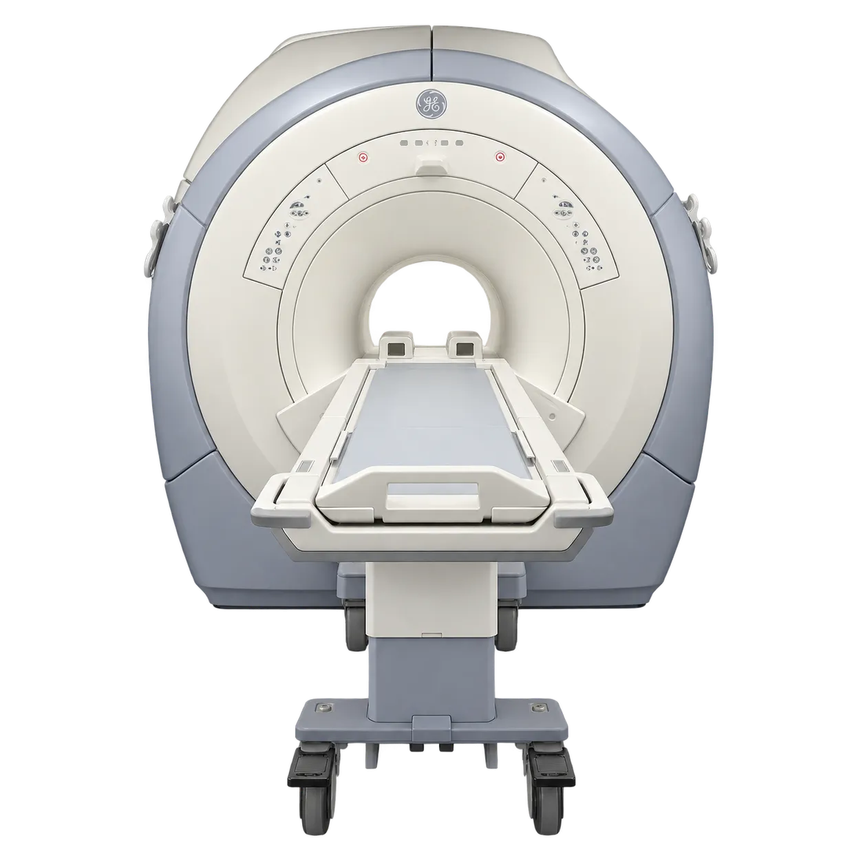
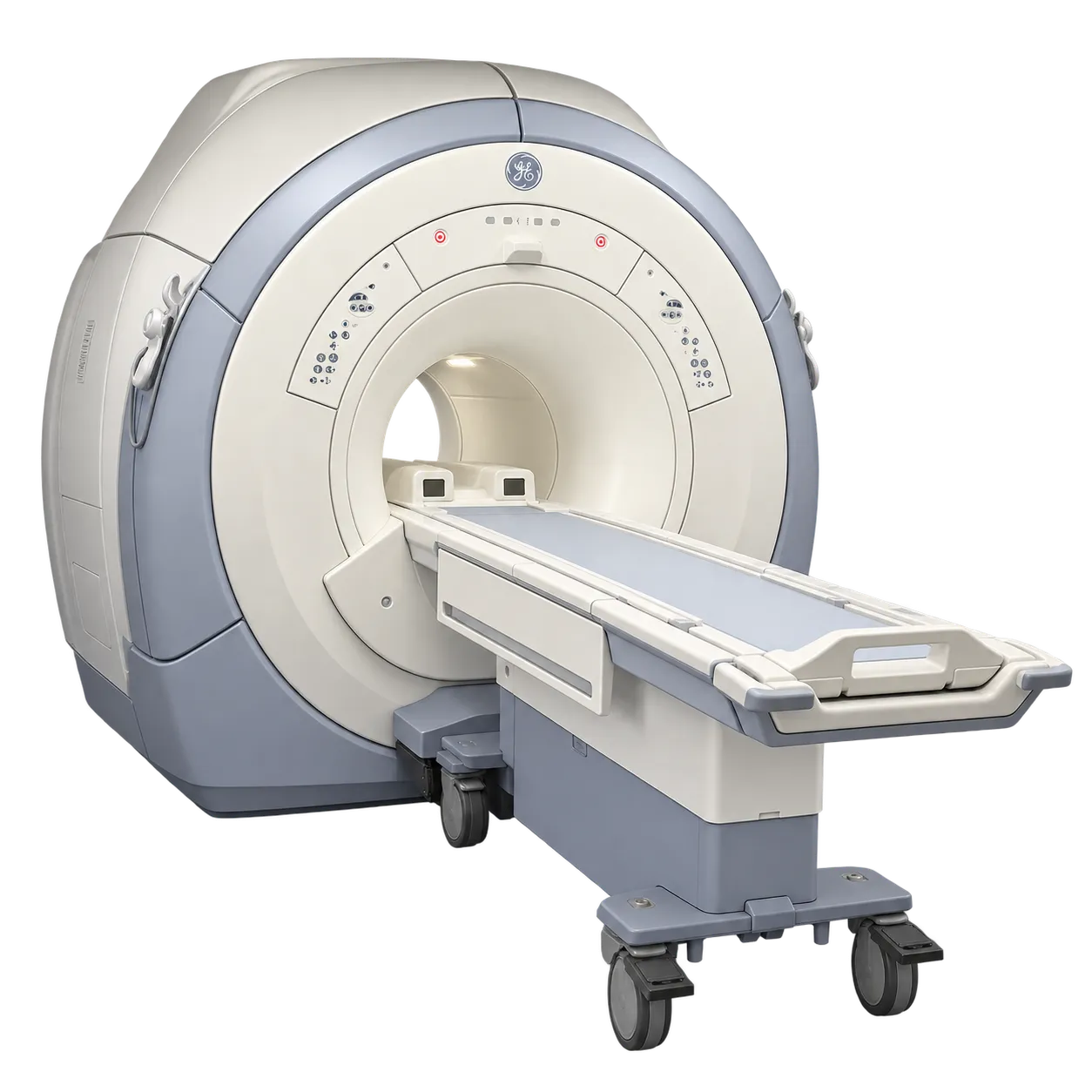
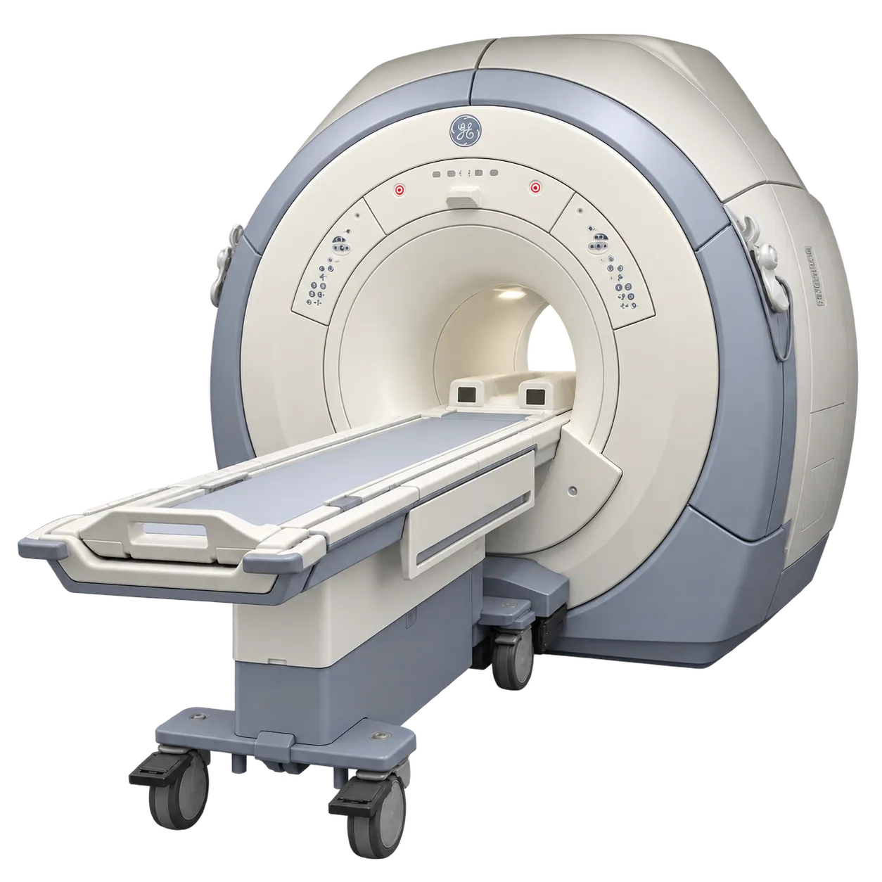
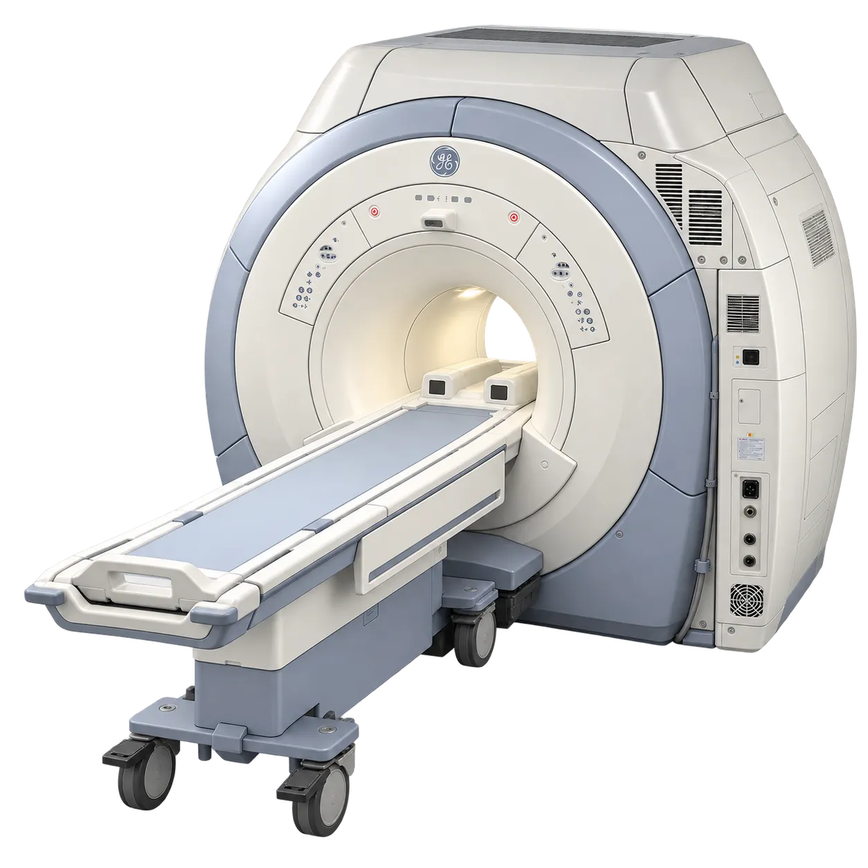
Departments with limited plant and room space are served by a single-cabinet configuration occupying approximately 25 square metres.
Services running long daily lists benefit from a detachable table, since preparation and transfer can overlap with scanning where a department is set up that way.