
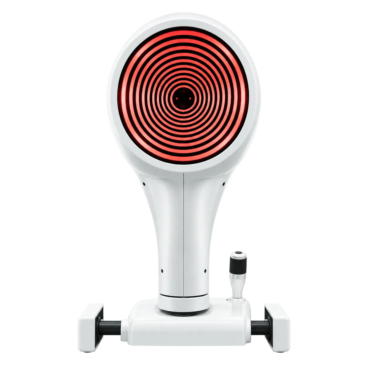
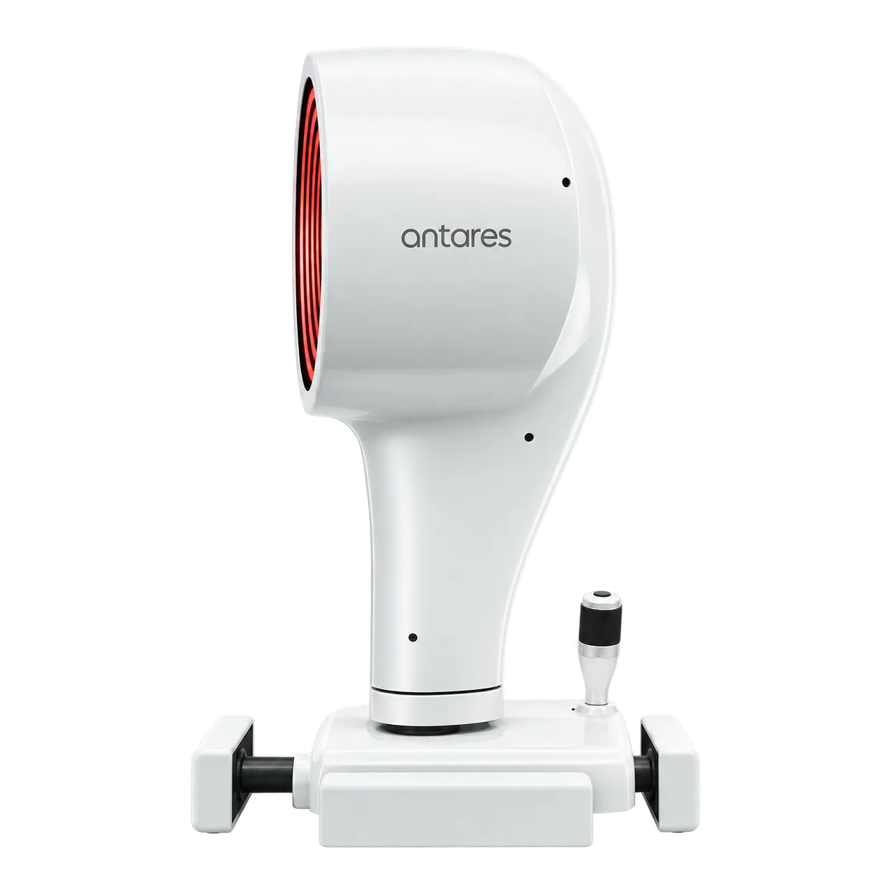
Optometry practices, contact lens clinics, dry eye clinics, and ophthalmology clinics use the Antares to examine the cornea and ocular surface. Optometrists, contact lens practitioners, ophthalmologists and ophthalmic technicians operate it, and the results inform dry eye assessment, contact lens fitting and screening before corneal surgery.
The sitting is built around one patient position. Placido topography, tear break-up time, the dry eye report, meibography and pupillography all run from the same instrument, so the patient stays at the chin rest while the operator changes analyses in software. Lighting changes with the task: white LEDs for topography and tear film, a 470 nm fluorescein stimulation LED for cobalt blue videokeratoscopy, and infrared for the meibomian glands and pupil.
For dry eye work, the report combines OSDI, eye redness, meibomian gland and tear meniscus findings with NIBUT, and the glands are imaged beside the corneal maps. Contact lens practitioners simulate rigid lens fits from an international lens database and use Autofit across more than 50,000 lenses. Keratoconus screening gives an ectasia-risk indication before corneal surgery, and pupillography at 0.04, 4 and 50 lux adds a dynamic record.
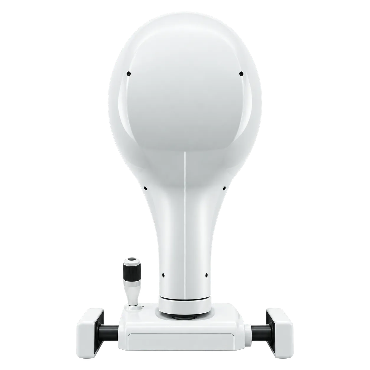
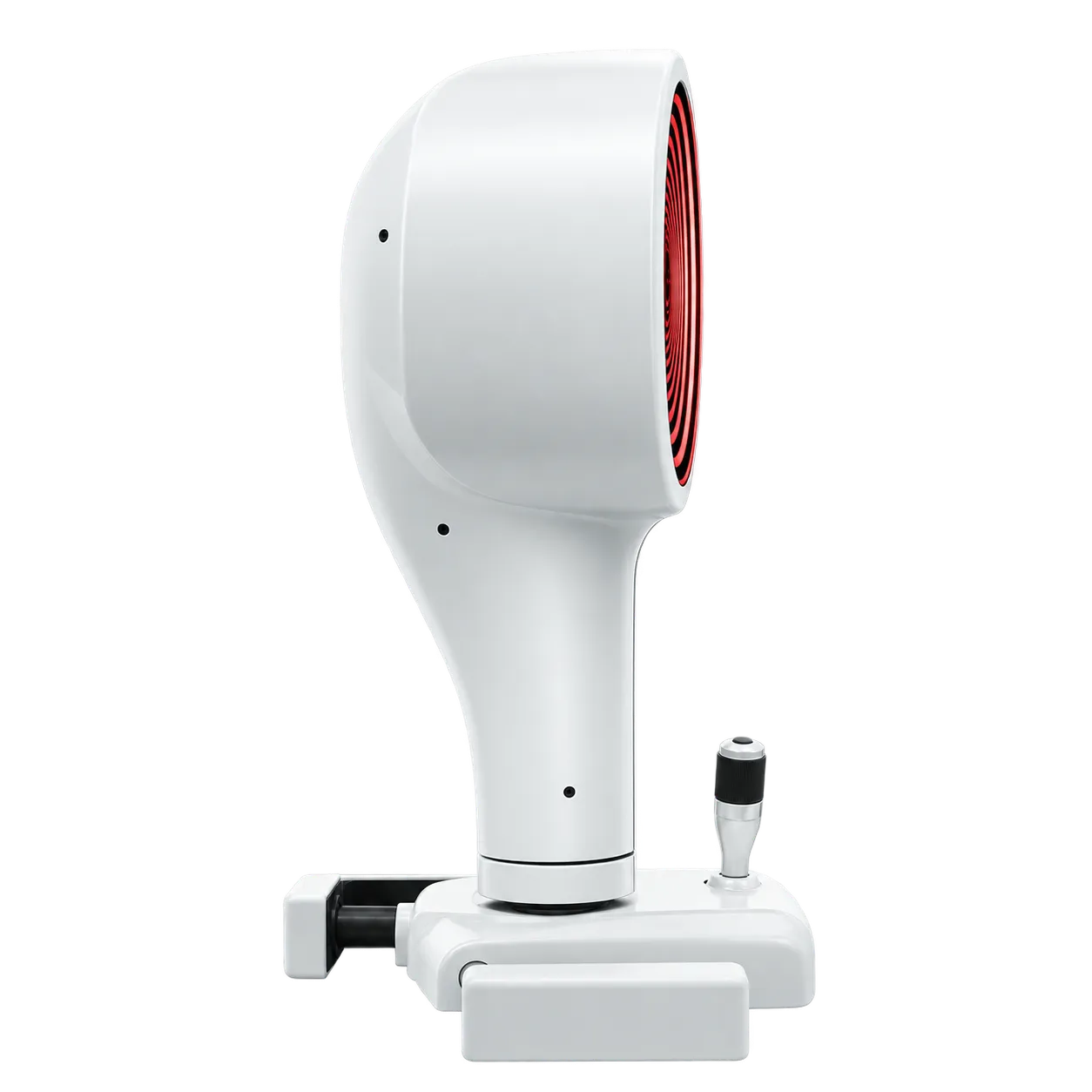
At 6.5 kg, with a 315 × 255 mm footprint, the head sits on a consulting-room table or an optional electric table. Data moves over USB 3.0 to a Windows 11 computer, and DICOM v3 compatibility under the IHE Eye Care profile allows examinations to join an imaging workflow. The same head serves a dry eye screening visit, a contact lens fitting appointment and a pre-surgical corneal check, so one instrument covers the routine work of a busy clinic day without a second device being needed.