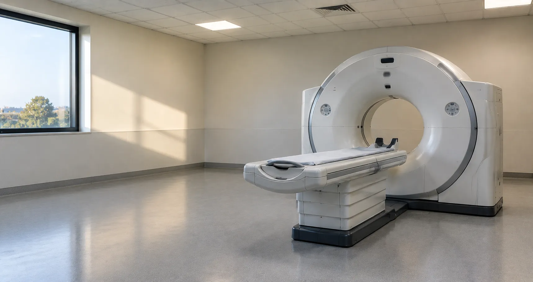
A cardiac CT scanner is a computed tomography system tuned to image the heart and coronary arteries, using a fast-rotating X-ray tube and detector array to build cross-sectional images precise enough to show the coronary lumen and calcified or soft plaque. The defining challenge it solves is motion: the heart never stops, so a cardiac CT scanner must acquire its images in a fraction of a heartbeat and synchronise them to the ECG. Coronary CT angiography, the main application, depends entirely on the scanner's ability to freeze that movement sharply.
For an imaging department or procurement lead, buying a cardiac CT scanner is a major capital decision where a handful of specifications,-temporal resolution, detector coverage, dose performance, and gating- determine whether coronary studies are diagnostic. It is also a regulated radiation installation, which shapes siting and running costs well beyond the purchase price.
Freezing a moving heart: temporal resolution and gating
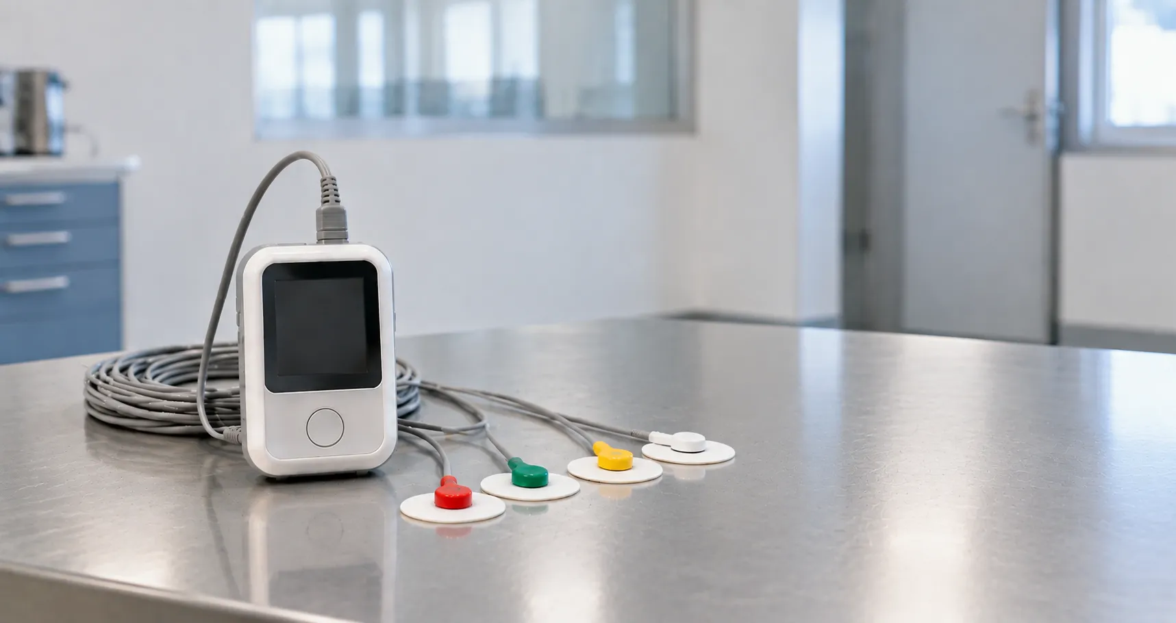
Temporal resolution, the effective time to capture one image, is the specification that most defines a cardiac CT scanner, because it decides how much cardiac motion blurs the coronaries. It is driven mainly by gantry rotation speed, with dual-source designs halving the effective acquisition time by using two tube-detector pairs. The scanner ties acquisition to the ECG through gating: prospective gating triggers the exposure only during the quiet phase of the cycle to save dose, while retrospective gating records continuously and reconstructs the best phase, useful for functional analysis. Getting sharp, motion-free coronary images depends on both fast rotation and reliable ECG synchronisation.
Detector coverage and rotation speed
The detector array's coverage along the patient decides how much of the heart is captured per rotation. Wide-detector systems can cover the whole heart in a single beat, removing the stitching artefacts that arise when a scan is assembled from several heartbeats and reducing sensitivity to rhythm variation. Narrower detectors acquire the heart over several beats using helical or step-and-shoot modes. Alongside coverage, rotation speed and the number of detector rows shape image quality and speed. When comparing cardiac CT scanners, weigh detector width, rotation time, and reconstruction power together, since they jointly determine performance on fast or irregular heart rates.
Radiation dose and dose-reduction features
Dose is a central specification for a cardiac CT scanner because coronary imaging must balance image quality against the radiation the patient receives. Modern scanners cut dose through prospective ECG-gated acquisition, automatic tube-current modulation, lower tube voltages for suitable patients, and iterative or deep-learning reconstruction that preserves image quality at lower exposure. Because these techniques directly affect both dose and diagnostic confidence, they are a real point of comparison between systems, not marketing detail. Any purchase must sit within a radiation service that monitors and optimises dose, so ask for documented dose performance on representative coronary protocols rather than a single headline figure.
Contrast, workflow and heart-rate control
Coronary CT angiography needs iodinated contrast timed to opacify the coronaries as the scan runs, which usually means an integrated or synchronised contrast injector and bolus-tracking software. Because image sharpness improves at lower, steadier heart rates, protocols often involve rate control before scanning, so the scanner's tolerance of higher and variable rates is clinically important. Throughput also depends on reconstruction speed and how smoothly the system moves from acquisition to a reviewed dataset. When specifying a cardiac CT scanner, consider the whole workflow, injector integration, post-processing for coronary analysis and calcium scoring, and how results reach the reporting system.
Calcium scoring and wider cardiac applications
A cardiac CT scanner does more than coronary angiography. A coronary artery calcium score, a low-dose non-contrast scan that quantifies calcified plaque, is a common study used to help stratify cardiovascular risk, and the same scanner supports it. Cardiac CT is also used to assess cardiac structures, bypass grafts and, increasingly, to plan structural interventions such as valve procedures where accurate measurement of the anatomy matters. When you specify a scanner, think about the full range of cardiac work it will carry, because the post-processing packages for calcium scoring, coronary analysis and structural planning are often licensed separately and belong in the budget.
Radiation law and device standards for cardiac CT
A cardiac CT scanner sits under two regulatory layers. As medical electrical equipment, it is built to IEC 60601-1, with the particular requirements for the safety of computed tomography X-ray equipment set out in IEC 60601-2-44. As a source of ionising radiation, it also falls under UK radiation law: the Ionising Radiations Regulations 2017, enforced for workplace radiation protection by the Health and Safety Executive, and the Ionising Radiation (Medical Exposure) Regulations 2017, or IR(ME)R, which govern justification and optimisation of patient exposures; guidance on both is published on GOV.UK. The device itself is regulated by the MHRA and must carry UKCA or CE marking. This guide draws on manufacturer documentation and UK regulatory guidance for procurement and clinical-engineering teams.
Siting, shielding and installation
A cardiac CT scanner is a fixed installation with substantial practical demands. The room needs radiation shielding designed and verified by a medical physics expert, adequate floor loading for a heavy gantry, controlled temperature and ventilation for the electronics, and space for the control area and contrast injector. Installation and commissioning include acceptance testing and dose verification before clinical use. These siting requirements, along with power and cooling, are a real part of the total cost and lead time, so estates, medical physics and radiology must be involved early. Underestimating the room works is a common way for a CT project to slip.
Specifying a cardiac CT scanner
Before committing to a cardiac CT scanner, assess the specifications and the installation together:
-
Temporal resolution and gantry rotation time, including whether a dual-source design is needed for high heart rates.
-
Detector coverage and row count, and whether whole-heart single-beat acquisition is required.
-
Dose-reduction toolkit: prospective gating, tube-current modulation, low kV and advanced reconstruction.
-
ECG gating options, plus tolerance of higher and irregular heart rates.
-
Contrast injector integration, bolus tracking, calcium scoring and coronary post-processing licences.
-
Compliance with IEC 60601-1 and IEC 60601-2-44, UKCA or CE marking, and IRR 2017 and IR(ME)R 2017 obligations.
-
Room shielding, floor loading, power, cooling, installation lead time and acceptance testing.
Reconstruction, workstation and reporting
A cardiac CT scanner produces large volumes of thin-slice data that need reconstructing and reviewing, so the reconstruction engine and cardiac workstation are as much a part of the purchase as the gantry. Coronary studies rely on tools that rebuild the vessels in multiple planes, straighten and measure the coronaries, and quantify calcium, and increasingly on advanced or AI-assisted reconstruction that lifts image quality at lower dose. Reconstruction speed affects how quickly a scanned patient becomes a reviewed dataset, which drives list throughput. Confirm what workstation licences and post-processing packages are included, how many concurrent users they support, and how images and reports flow into your PACS and reporting system.
Service contracts, tube life and running costs
The purchase price of a cardiac CT scanner is only part of the commitment. The X-ray tube is a major wear item with a finite lifespan and a significant replacement cost, so its expected life and warranty terms belong in any comparison. Ongoing costs include the service contract, planned maintenance, dose and quality-assurance testing, software and reconstruction licences, and contrast consumables. A full-service agreement with defined uptime and response times protects a busy coronary CT list. Balance capital price against these lifetime costs when comparing systems; MediGear helps UK facilities compare verified imaging suppliers and service terms through the buyers page or current suppliers.
Choosing well for a coronary CT service
A cardiac CT scanner lives or dies on its ability to freeze a moving heart, so temporal resolution, detector coverage, gating and dose performance are the specifications that decide whether coronary studies are diagnostic. Around those sit the practical realities: contrast workflow, room shielding, radiation compliance and the tube and service costs that run for the machine's life. Weigh the imaging performance against the installation and lifetime running costs together, and the cardiac CT scanner will support a reliable, well-optimised coronary imaging service.
Disclaimer
This article is for informational purposes only. It is published by MediGear (medigear.uk) for general information and procurement guidance, and is not clinical, diagnostic, treatment, technical, engineering, legal or regulatory advice, nor a product endorsement, guarantee or substitute for professional assessment. MediGear does not provide medical consultations. Buyers should consult their clinical, biomedical, estates and regulatory contacts, and the manufacturer's documentation, and independently verify all specifications, certifications, compatibility and suitability before purchase. Specifications, certifications and availability are correct at the time of publication and may change without notice. MediGear is a medical-equipment distributor and does not sell medicines or pharmaceutical products.