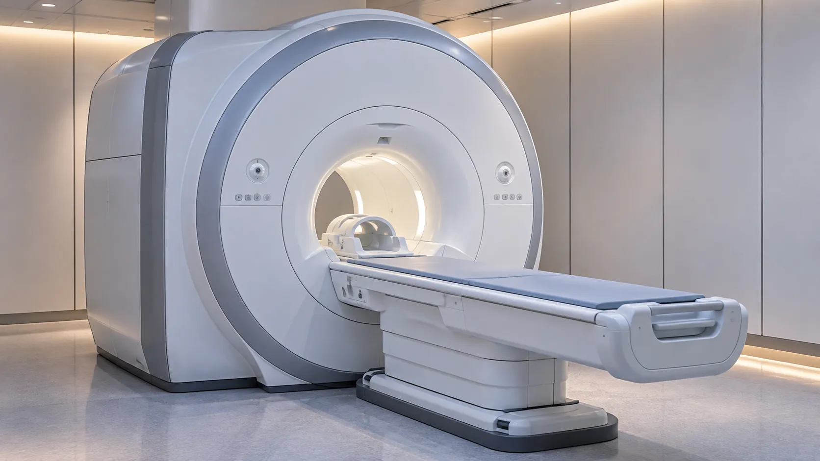
Cardiac MRI, also called cardiovascular magnetic resonance, images the heart using a strong magnetic field and radio waves rather than ionising radiation, so a department can assess chamber size, wall motion, valve flow and the state of the heart muscle itself in one sitting. It is the reference method for measuring ventricular volumes and ejection fraction, and the only routine scan that characterises the myocardium — separating scar from inflammation from infiltration — without a biopsy.
For an imaging lead or procurement team, a cardiac MRI system is one of the largest capital and estates commitments a diagnostic service makes. The clinical value is high, but so is the siting, shielding and cryogen burden. A handful of choices — field strength, coils, gating and analysis software — decide whether the heart studies are quick and diagnostic or slow and marginal.
Why departments add cardiac MRI to a heart-imaging service
Echocardiography is faster and cheaper for first-line work, and cardiac CT excels at the coronary arteries. Cardiac MRI earns its place where the tissue matters: telling infarct scar from viable muscle, spotting myocarditis, quantifying fibrosis, and measuring volumes reproducibly enough to track a patient over years. Because it uses no X-rays, it suits younger patients and repeat imaging where cumulative dose would be a concern. The National Institute for Health and Care Excellence sets out where cardiovascular magnetic resonance is indicated in its clinical guidance at nice.org.uk, and matching your caseload to those pathways is the first step in justifying the spend.
This guide is compiled from manufacturer specifications, the IEC 60601 standards family and current UK regulatory guidance, and is written for procurement and clinical-engineering teams rather than patients. MediGear is a UK medical-equipment distributor that helps facilities compare verified suppliers on like-for-like terms.
Structure, function and tissue: what a cardiac MRI scan shows
A cardiac MRI study is really several sequences stitched into one protocol. Cine imaging, usually a balanced steady-state free precession sequence, gives moving pictures of the beating heart for wall motion, volumes and ejection fraction. Late gadolinium enhancement highlights scar and fibrosis, which hold contrast differently from healthy muscle. Parametric mapping — T1, T2 and extracellular volume — puts a number on tissue properties, catching diffuse fibrosis, oedema and infiltration that older sequences missed. Phase-contrast sequences measure blood-flow velocity across valves. The buyer's takeaway is simple: the scanner is only as capable as the sequences and coils it ships with, so the software licence matters as much as the magnet. A system sold cheaply but stripped of mapping or flow sequences will not deliver a full cardiac service, and adding them later is rarely cheap.
Field strength and the magnet at the centre of the system
Most cardiac MRI runs at 1.5 tesla, which balances image quality, throughput and a mature set of cardiac sequences. Systems at 3 tesla give a higher signal that can sharpen some work but bring more artefacts around the heart and lungs, so the higher field is not automatically better for cardiac use. The superconducting magnet is cooled by liquid helium and stays energised continuously. The gradient coils that encode spatial information and the radiofrequency coils that receive signal matter just as much to image quality. Dedicated cardiac phased-array surface coils with enough channels are essential for fast parallel imaging, so confirm the coil set is specified for cardiac work rather than bundled as general-purpose.
Gating and breath-holds: imaging a target that never stops
The heart moves twice — it beats, and it rides on the breathing cycle — and a cardiac MRI system has to freeze both. ECG gating synchronises acquisition to the cardiac cycle so each image is built from the same phase across many beats. Respiratory compensation, whether breath-holding or navigator-based free-breathing, handles the slower motion. Newer accelerated and free-breathing sequences shorten scans and rescue patients who cannot hold their breath or who have an irregular rhythm. Reliable gating hardware and a strong arrhythmia-rejection algorithm are what separate a smooth cardiac list from a frustrating one.
The MR safety zone your estate has to build
An MRI is never just a machine in a room. The magnet stays on permanently, so the suite needs a controlled four-zone layout, a radiofrequency-shielded Faraday cage, a quench pipe to vent helium safely, ferromagnetic screening, and strict access control. Projectile incidents and implant compatibility are the dominant safety risks, which is why the Medicines and Healthcare products Regulatory Agency publishes detailed guidance on the safe use of MRI equipment at mhra.gov.uk. Build in the staffing for an MR safety officer and the workplace duties enforced by the Health and Safety Executive at hse.gov.uk before the scanner arrives, because retrofitting a compliant suite is expensive.
Cardiac MRI standards and compliance
As medical electrical equipment, an MRI scanner is built to the general standard IEC 60601-1 and the MR-specific particular standard IEC 60601-2-33, which sets the magnetic, gradient and radiofrequency exposure limits. The device must carry UKCA or CE marking under MHRA oversight. Unlike CT or nuclear studies, cardiac MRI uses no ionising radiation, so the Ionising Radiations Regulations do not apply — but the static-field, gradient and RF controls very much do, alongside the site's own MR safety governance. Ask to see the scanner's conformance documentation and the site's safety framework as one package.
Contrast, gadolinium and going contrast-free
Gadolinium-based contrast agents make late gadolinium enhancement possible and remain central to scar imaging. They bring their own procurement and governance strands: agent choice, cannulation workflow and renal-function checks handled by the clinical team. The shift toward parametric mapping means some tissue questions can now be answered without contrast at all, which trims consumable cost and helps patients who cannot receive gadolinium. A scanner shipped with validated mapping sequences future-proofs the service.
Bore, throughput and patient comfort
Bore diameter and table design affect who you can scan and how quickly the list moves. A wider bore eases larger or claustrophobic patients and speeds positioning, while quieter gradient modes improve tolerance. Throughput on a cardiac list is governed less by the magnet than by protocol length, coil setup,p and reporting workflow, so weigh accelerated sequences and slick patient handling when you compare systems.
Running costs beyond the magnet
The purchase price is only the start. A superconducting cardiac MRI system consumes helium and relies on a cold head and compressor to limit boil-off; a failed cold head can end in a costly quench. Budget for the service contract, coil repair or replacement — coils are delicate and expensive — software upgrades, and the power and cooling the magnet room draws around the clock. When you compare quotes, compare full lifetime cost, and use MediGear's tools to weigh suppliers rather than headline price alone.
Adding stress perfusion to a cardiac MRI service
Beyond structure and tissue, cardiac MRI can assess perfusion under stress. A vasodilator such as adenosine is given while first-pass contrast sequences watch blood reach the muscle, revealing territories that are underperfused when demand rises. Stress cardiac MRI competes with nuclear perfusion imaging but adds no ionising radiation, and it pairs perfusion with the same scan's scar and function data. If your service intends to offer stress work, confirm the scanner ships with validated perfusion sequences, the monitoring to run a pharmacological stress safely, and the staffing model to support it.
Reporting, analysis and integration
A cardiac MRI study generates large, multi-sequence datasets that need dedicated post-processing: contouring the ventricles for volumes and ejection fraction, quantifying mapping values, and measuring flow. The analysis package and its licences shape how quickly a reporter turns a scan around, so treat software as a first-order specification, not a bundled extra. Confirm the system exports cleanly to your PACS, links to the cardiology information system, and supports the structured reporting your department uses. Integration failures are a common, avoidable drag on throughput.
Specifying a cardiac MRI system: a buyer's checklist
-
Field strength (1.5T versus 3T) matched to your cardiac caseload, not just headline resolution.
-
Dedicated cardiac phased-array coils with enough channels for parallel imaging.
-
Cine, late gadolinium enhancement, T1/T2 and extracellular-volume mapping, ng and phase-contrast flow sequences, licensed and included.
-
ECG gating quality and arrhythmia rejection, plus free-breathing options for difficult patients.
-
Conformance to IEC 60601-2-33 and current UKCA or CE marking.
-
Site readiness: Faraday cage, quench pipe, four-zone layout and named MR safety governance.
-
Cryogen and cold-head arrangement, service-contract cover and coil-replacement terms.
-
Post-processing and reporting software, plus PACS and cardiology-system integration.
Choosing well for your imaging service
A cardiac MRI purchase is a decade-long commitment, so judge it on the whole picture: the sequences and coils that make heart studies diagnostic, the estates work the magnet demands, and the running costs that follow the invoice. Get the specification right, and the scanner will carry structural, functional,l and tissue imaging that no other single modality matches. If you are scoping a system, MediGear can help you reach the right suppliers and compare them on equal terms.
Disclaimer
This article is for informational purposes only. It is published by MediGear (medigear.uk) for general information and procurement guidance, and is not clinical, diagnostic, treatment, technical, engineering, legal or regulatory advice, nor a product endorsement, guarantee or substitute for professional assessment. MediGear does not provide medical consultations. Buyers should consult their clinical, biomedical, estates and regulatory contacts, and the manufacturer's documentation, and independently verify all specifications, certifications, compatibility and suitability before purchase. Specifications, certifications and availability are correct at the time of publication and may change without notice. MediGear is a medical-equipment distributor and does not sell medicines or pharmaceutical products.