
Introduction
Echocardiography uses ultrasound to show the heart as it beats — its chambers, valves, walls and blood flow — without any radiation. It is one of the most widely used cardiac investigations, guiding decisions from a first breathless clinic visit to a complex valve operation. Because the pictures are created and interpreted in real time, the quality of the machine and the probe matters enormously, and so does the software that measures and reports what the operator sees.
Equipping an echocardiography service spans the echo machines themselves, the range of probes for surface and swallowed imaging, provision for stress and contrast studies, and the reporting and measurement tools that turn images into clinical answers. This echocardiography equipment buyer guide outlines what each area needs, how the pieces fit together, and the quality and traceability questions to settle early on. It is written for cardiologists, cardiac physiologists, sonographers, and procurement professionals across the UK, Europe, the US, the Gulf, and South Africa.
What an echo service needs — the short version
-
Echocardiography machines matched to the setting, from full cardiology systems to portable units.
-
A range of probes — transthoracic and transoesophageal — for surface and detailed imaging.
-
Stress and contrast capability, with reporting software to measure and record findings.
Start with the echo machine and probes.s
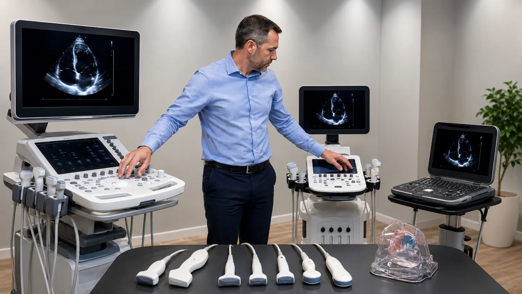
The core purchase is the echocardiography machine and the accompanying probes. A full cardiology system offers the image quality, Doppler modes, and measurement tools that a specialist service relies on. At the same time, smaller portable and handheld units extend imaging to the ward, clinic, and bedside. The choice matches where studies are performed and how demanding they are, because a busy echo laboratory and a point-of-care setting have very different needs.
Probes shape what the service can image. A transthoracic probe placed on the chest covers most studies, while a transoesophageal probe, passed into the gullet, gives detailed views used in valve assessment and during surgery. Once machines and probes are settled, the service adds stress and contrast capability, plus reporting software that measures function and stores results, so a study flows from image to report without gaps.
What echocardiography needs
-
Cardiology echo system. A full-feature machine with the Doppler modes and measurements a specialist service needs.
-
Portable and handheld units. Smaller systems for ward, clinic and point-of-care imaging.
-
Transthoracic probes. Surface probes covering the majority of routine studies.
-
Transoesophageal probe. Detailed imaging for valves and intraoperative use.
-
Stress echo capability. Provision for exercise or pharmacological stress imaging.
-
Contrast echo support—enhancement for difficult-to-image patients, where used.
-
Reporting and measurement. Software to quantify function and produce structured reports.
-
Probe care and storage: cleaning, disinfection and safe storage for delicate probes.
The equipment checklist
|
Equipment |
Role in the service |
Key selection point |
|
Cardiology echo system |
Core imaging |
Image quality; Doppler; measurements |
|
Portable echo unit |
Ward and clinic imaging |
Portability; image quality |
|
Transthoracic probe |
Routine studies |
Frequency range; ergonomics |
|
Transoesophageal probe |
Detailed and intraoperative views |
Compatibility; reprocessing |
|
Stress echo provision |
Functional assessment |
Protocol support; workflow |
|
Reporting software |
Measurement and reporting |
Quantification; storage link |
|
Probe reprocessing |
Infection control |
Validated cleaning; storage |
Transthoracic and transoesophageal imaging
Most echocardiograms are transthoracic, taken with a probe on the chest, and this is where a service does the bulk of its work. Transoesophageal imaging is more specialised: a probe is passed into the oesophagus, positioned close behind the heart, and produces detailed images of the valves and structures that are hard to see from the surface. It is central to valve assessment and to guiding cardiac surgery and interventions. Planning for both, with the probe care and reprocessing they demand, lets a service cover routine and complex work alike.
Stress and contrast echo
Echocardiography does more than image the resting heart. Stress echo compares the heart at rest with the heart under exercise or pharmacological stress to reveal problems that only appear on exertion. At the same time, contrast agents can sharpen the picture in patients who are difficult to image. Both extend what the service can answer and shape equipment planning—stress protocols need the right software and supporting kit, and contrast work needs its own supplies and preparation. Deciding which to offer is part of matching the service to its caseload.
Compliance and documentation
Echocardiography equipment must be genuine, current and fully documented. In the UK, the MHRA regulates medical devices, and NICE publishes clinical guidance. The ISO maintains international standards for medical devices. Requirements vary by country and market, so confirm current obligations with the relevant authorities before you buy.
Support keeps an echo service running.
-
Installation, commissioning and clinical familiarisation for machines and probes.
-
Preventive maintenance and probe testing that protect image quality and patient safety.
-
Reliable supply of probes, contrast supplies, electrodes and consumables.
-
Rapid technical response is essential, because a downed machine or probe stalls a clinic list.
-
Clear documentation and traceability for machines, probes and reprocessing records.
More from Business
-
Buyers — source compliant, fully documented equipment.
-
Suppliers — list your equipment and reach buyers worldwide.
-
Advertise — promote your products and services to a clinical audience.
-
Business overview — explore everything MediGear offers healthcare organisations.
Disclaimer
This article is for general information only. It does not constitute clinical, procurement, legal or regulatory advice. Equipment requirements for echocardiography vary by service, setting, facility, country and market, and change over time. Always confirm current requirements with the relevant authorities and consult qualified clinical, physiological, biomedical engineering, procurement, and regulatory professionals for decisions specific to your organisation. medigear.uk accepts no liability for actions taken based on this content.
"The heart, seen clearly." An echo service equipped for every study.
We supply medical equipment to healthcare organisations across the UK, Europe, the US, the Gulf and South Africa. Everything is genuine, documented and supported, so your echocardiography service images with confidence.
Frequently Asked Questions
An echo service needs echocardiography machines matched to its needs—full cardiology systems for the laboratory and portable or handheld units for ward and clinic imaging—plus a range of probes. Transthoracic probes are used for routine studies, and a transoesophageal probe provides detailed valve and intraoperative views. Most services also need stress- and contrast-capability reporting and measurement software, and validated probe reprocessing and storage.
Transthoracic echocardiography uses a probe placed on the chest and is used in most studies. Transoesophageal echocardiography passes a probe into the oesophagus, where it sits close behind the heart and produces detailed images of the valves and structures that are hard to see from the surface. It is used to assess valves and guide cardiac surgery and interventions, and it needs careful reprocessing between patients.
Stress echo compares the heart at rest and under stress — either exercise or a medication that mimics it — to reveal problems, such as reduced blood supply, that only appear on exertion. It needs the right protocol support and software, along with the echo machine. Offering it broadens what a service can answer, so it is planned as part of matching equipment to the expected caseload.
Yes. Full cardiology systems provide the image quality, Doppler modes and measurement tools that a specialist laboratory relies on. At the same time, portable and handheld units bring imaging to the ward, clinic and bedside. Many services run both, matching the machine to where and how the study is performed. Point-of-care imaging and a busy echo laboratory have quite different requirements.
Yes. Machines, probes and reprocessing should be genuine, current and traceable. In the UK, the MHRA regulates devices, NICE provides clinical guidance, and ISO maintains international device standards. Requirements vary by country and market, so confirm current obligations with the relevant authorities before purchasing.
Yes. MediGear supplies medical equipment to healthcare organisations across the UK, Europe, the US, the Gulf and South Africa. As a distributor, we provide genuine, documented and supported equipment, from cardiology echo systems and probes to portable units and reporting software. Requirements differ by service and market, so we work with your team to match equipment to your service and its regulatory setting.