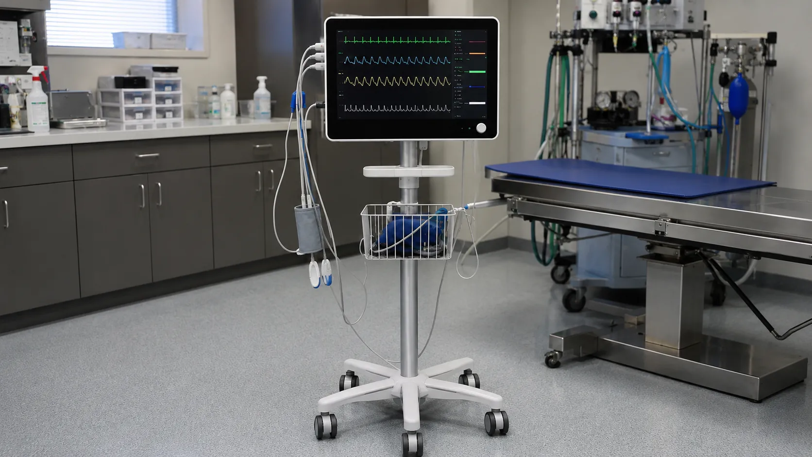
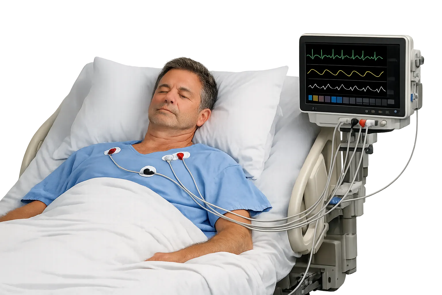
Impedance pneumography measures breathing by tracking the tiny changes in electrical resistance across the chest as the lungs fill and empty. As air enters the thorax, the chest becomes slightly less conductive, and as the patient exhales, conductivity rises again; a monitor reads that rhythmic change and reports it as a respiratory rate and a breathing waveform. It is the method most bedside patient monitors use to count breaths, because it reuses the same electrodes already placed for the ECG. This guide explains the measurement principle and what a procurement lead should weigh when specifying monitors that rely on it.
The technique is popular precisely because it adds respiratory monitoring at almost no extra hardware cost. No mask, flow sensor or separate probe is needed, which is why it is built into so many multiparameter monitors and neonatal apnoea systems.
The measurement principle
A very small, high-frequency alternating current is passed between two of the ECG electrodes on the chest, well below the level a patient can feel. The monitor measures the voltage needed to drive that current, which reflects the electrical impedance of the tissue between the electrodes. Because air is a poor conductor, thoracic impedance rises during inspiration and falls during expiration. The monitor extracts this slow rise and fall from the signal, filters out the faster cardiac component, and counts the cycles to produce breaths per minute alongside a respiration waveform.
Electrodes and lead configuration
Impedance pneumography shares the ECG electrodes, so lead placement affects the quality of the breathing signal as much as the heart trace. The respiration measurement is usually taken across the lead axis that best captures chest-wall movement, commonly between the right-arm and left-arm electrodes. Poor electrode contact, dried gel or placement over bone rather than moving tissue all weaken the signal. Because the same electrodes serve two purposes, buyers should confirm that a monitor lets clinicians select or optimise the respiration lead independently of the displayed ECG lead.
Reading the respiratory waveform
The output is a respiration trace whose amplitude reflects the depth of chest movement and whose frequency gives the rate. A steady, well-formed waveform allows the monitor to set apnoea alarms when breathing pauses beyond a set interval. The waveform is a relative signal rather than a calibrated tidal volume, so it shows the pattern and timing of breathing rather than the exact volume of air moved. That distinction matters when comparing it with methods such as spirometry or capnography that quantify gas movement directly.
Known limitations to design around
No respiratory method is perfect, and understanding the weaknesses of impedance pneumography prevents over-reliance on it.
Cardiogenic artefact
The beating heart also changes thoracic impedance, so the pulse can be mistaken for a breath. This cardiac interference can cause a monitor to overcount during genuine apnoea, which is a recognised limitation in neonatal apnoea monitoring. Good signal processing and correct lead placement reduce, but do not entirely remove, the effect.
Motion and obstructive events
Patient movement, coughing or handling produces impedance changes that mimic breaths, and the method detects chest-wall motion rather than airflow, so it can register effort during an obstructed breath where no air actually moves. For patients where airflow itself must be confirmed, capnography or a dedicated flow sensor is the more appropriate tool, and many buyers specify both.
Where impedance pneumography is used
The method appears across a wide range of care settings. It is standard on general-ward and high-dependency multiparameter monitors, on transport monitors, and in neonatal and paediatric units where continuous apnoea surveillance is needed. In lower-acuity or domiciliary settings it underpins simple respiratory-rate monitoring without the cost of a gas-analysis front end. Matching the monitor to the setting means deciding how much you need airflow confirmation, which points some critical and anaesthetic areas towards adding capnography rather than relying on impedance alone.
How it compares with other respiratory methods
Impedance pneumography is one of several ways to monitor breathing, and choosing well means knowing the alternatives. Capnography measures exhaled carbon dioxide and confirms that air is actually moving, which impedance cannot do, making it the reference where airflow must be proven, such as during procedural sedation. A dedicated flow sensor or spirometry quantifies tidal volume directly but needs a mask or airway connection the patient may not tolerate. Acoustic and accelerometer-based respiration sensors offer further alternatives that avoid electrode dependence. Against these, impedance pneumography wins on simplicity and cost, since it piggybacks on the ECG electrodes already in place and gives a continuous rate and waveform with no extra patient interface. The sensible position for most buyers is to treat impedance as the default ward method while specifying capnography for the specific areas where confirming airflow is clinically necessary.
Getting a clean respiration signal in practice
The quality of an impedance respiration trace rests heavily on how electrodes are applied, so buying good monitors is only half the job. Electrodes should sit on moving chest-wall tissue rather than over bone, with fresh gel and firm skin contact, and the respiration lead should be chosen to capture the largest breathing swing. Excess body hair, sweat and patient position all degrade the signal, and staff need to know how to reposition electrodes when the trace deteriorates. Because the same electrodes drive the ECG, a compromise that suits the heart trace may weaken the respiration signal, so monitors that let the two be optimised separately are worth paying for. Training ward staff on placement pays back in fewer false apnoea alarms and more reliable rate counting, which is a workflow benefit as much as a technical one.
Specifications a buyer should check
When comparing monitors, look past the fact that they all claim respiration monitoring. Check the respiratory-rate range and how low it reads for neonates, the apnoea-detection delay and how it is set, and the quality of the artefact rejection, particularly cardiac-overcounting suppression. Ask whether the respiration lead can be selected separately, how the alarms escalate, and whether the monitor can display capnography as an option for areas that need airflow confirmation. Confirm the electrode types and connectors are ones you already stock, because an incompatible lead set adds a hidden consumable cost.
Standards, calibration and consumables
Multiparameter monitors supplied in the UK should carry UKCA or CE marking appropriate to their class and are built to the IEC 60601 family for medical electrical safety, with the particular standard for respiration monitoring applying to the breathing function. Impedance pneumography needs no user gas calibration, but the monitor still follows a planned maintenance and electrical-safety testing schedule. The recurring cost sits in the ECG electrodes, since signal quality depends on fresh, well-adhered electrodes, so factor electrode consumption into the running cost. The MHRA regulates these monitors, and manufacturer documentation should state the respiration accuracy and intended use.
Procurement checklist
-
Confirm UKCA or CE marking and the IEC 60601 particular standard for the respiration function.
-
Check the respiratory-rate range, low-rate performance for neonates and apnoea-detection settings.
-
Assess artefact rejection, especially suppression of cardiac overcounting during apnoea.
-
Verify the respiration lead can be optimised independently of the displayed ECG lead.
-
Decide whether capnography is needed alongside impedance for airflow confirmation.
-
Confirm electrode types and connectors match your existing stock and forecast their use.
-
Review planned maintenance, electrical-safety testing and spare-part availability.
Conclusion
Impedance pneumography gives a low-cost, electrode-shared way to monitor breathing, and it suits most ward and transport settings well, provided buyers understand its blind spots around cardiac artefact and airflow. Knowing where it needs backing up with capnography is the key to specifying the right monitor. MediGear helps facilities compare patient monitors and their consumables across suppliers; register as a buyer or contact the MediGear team to match a monitor to your clinical areas.
Disclaimer
This article is for informational purposes only. It is published by MediGear (medigear.uk) for general information and procurement guidance, and is not clinical, diagnostic, treatment, technical, engineering, legal or regulatory advice, nor a product endorsement, guarantee or substitute for professional assessment. MediGear does not provide medical consultations. Buyers should consult their clinical, biomedical, estates and regulatory contacts, and the manufacturer's documentation, and independently verify all specifications, certifications, compatibility and suitability before purchase. Specifications, certifications and availability are correct at the time of publication and may change without notice. MediGear is a medical-equipment distributor and does not sell medicines or pharmaceutical products.