Depth of anaesthesia describes how strongly a patient is anaesthetised at a given moment — from light sedation, where awareness is still possible, through to the deep unconsciousness needed for major surgery. Because an anaesthetised patient cannot report their own experience, clinicians infer depth from a combination of clinical signs and, increasingly, monitors that process the brain’s electrical activity. This guide gives a broad overview of the range of methods used to judge depth today, rather than focusing on any single proprietary index.
Getting depth right matters in two directions. Too light risks accidental awareness during surgery, a rare but serious event; too deep is linked with slower recovery and other harms. The methods below span traditional bedside observation and the electronic monitors now common in theatres.
Why measuring depth is difficult.
There is no single, direct measure of consciousness. Anaesthesia is not one effect but several — hypnosis (unconsciousness), analgesia (pain control) and immobility — often produced by different drugs acting together. A patient can be deeply unconscious yet show a stress response, or be still simply because a muscle relaxant has removed the ability to move. This is why depth is inferred from several signals rather than read off one dial, and why no method is treated as infallible.
Clinical signs still form the baseline.
Long before electronic monitors, anaesthetists judged depth from the body’s responses, and these signs remain the foundation. Rising blood pressure and heart rate, sweating, tears, pupil changes, and movement or grimacing all suggest anaesthesia is lightening. Breathing pattern helps in a spontaneously breathing patient. Older structured scores combined such signs into a rough index. The major limitation is that muscle relaxants remove movement and that these autonomic signs are influenced by drugs and disease, so they can mislead when used alone.
End-tidal agent monitoring and MAC
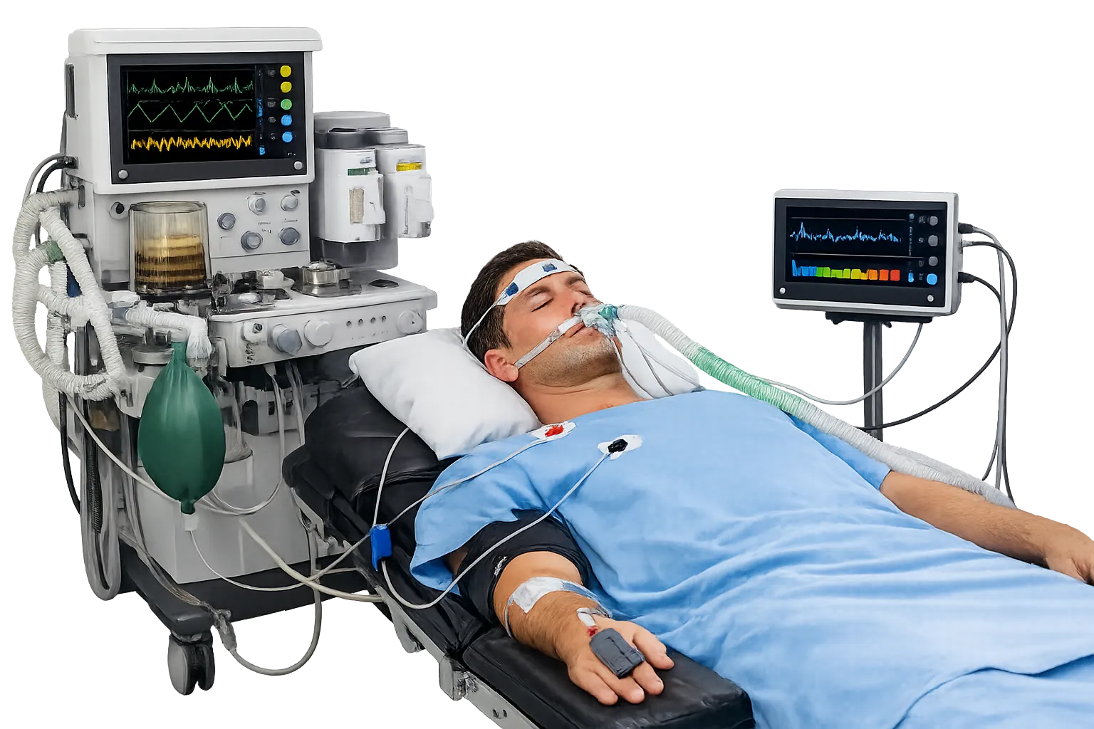
For inhaled anaesthesia, the concentration of volatile agent breathed out gives a useful surrogate for depth. Anaesthetic machines measure end-tidal agent concentration, which is compared against the minimum alveolar concentration (MAC) — the amount of agent needed to prevent movement in response to a surgical stimulus in half of patients. Tracking end-tidal agent as multiples of MAC helps keep a patient adequately but not excessively anaesthetised. MAC values differ between agents and are modified by age, temperature and other drugs, so the figure is a guide rather than a fixed target. This surrogate does not exist for intravenous-only anaesthesia, where the drug is delivered into the bloodstream rather than exhaled and cannot be sampled in the breath, which is a large part of why brain monitoring has drawn interest for those techniques.
EEG-based depth indices in general
The most widely used electronic monitors are based on processed electroencephalography. A sensor on the forehead captures the brain’s cortical electrical activity, which changes characteristically as anaesthesia deepens: fast, low-amplitude activity when awake gives way to slower, larger waves and, very deep, periods of suppression. An algorithm reduces this complex waveform to a single dimensionless number, usually scaled roughly from fully awake to deeply suppressed. Several commercial systems apply this principle with different algorithms; the shared idea is turning raw EEG into an easy-to-read trend.
Evoked potentials
A different approach measures the brain’s response to a repeated stimulus. Auditory evoked potentials, for example, record the electrical response to a series of clicks; the timing and size of the mid-latency response change predictably as anaesthesia deepens. Because it reflects how the brain processes a signal rather than only its background activity, this method offers a complementary view of depth. It appears in some commercial monitors and remains an active area of research and refinement. Because it captures a different aspect of brain function from the background activity that most indices summarise, it is sometimes used alongside them rather than as a replacement.
The isolated forearm technique
A classic reference method deserves mention. In the isolated forearm technique, a tourniquet is inflated on one arm before a muscle relaxant is given, so blood-borne relaxant cannot reach that hand. The patient can then move the hand to command if consciously aware, even while paralysed elsewhere. It is used mainly in awareness research rather than routine theatre work, but it underlines why movement alone is an unreliable guide once relaxants are in use.
What good depth control looks like in practice
In routine use, an anaesthetist aims to keep the patient adequately but not excessively anaesthetised, adjusting drug delivery in response to surgical stimulation and the signals available. The three components — hypnosis, analgesia and immobility — are balanced rather than driven by one measure, because a monitor showing adequate hypnosis says nothing about whether pain is controlled. Depth also varies between patients: age, frailty, other medication and illness all shift how much agent a given person needs, so fixed doses are a starting point rather than an answer. Titrating to the individual, guided by signs, agent concentration and any brain monitor, is what keeps a patient safely on the intended plane and supports a smoother recovery.
Limitations and clinical guidance
No depth measure is perfect. Processed indices can be affected by the specific drugs used, by electrical interference from muscle activity, and by individual variation, so they are interpreted alongside clinical signs and agent concentrations, not instead of them. UK guidance, including assessments from NICE, has considered depth-of-anaesthesia monitors particularly where the risk of awareness is higher — for instance during total intravenous anaesthesia combined with muscle relaxation, where agent monitoring cannot help. Local policy should define when such monitoring is used.
Where depth monitoring is used
Brain-based depth monitoring is not needed for every anaesthetic, and buyers should size purchases to where it earns its place. It is most often considered for total intravenous anaesthesia, especially when combined with muscle relaxants, and for patients thought to be at higher risk of awareness. It is also used to guide sedation depth in some intensive-care settings and can help avoid unnecessarily deep anaesthesia in frail or elderly patients, who may be more sensitive to agents. General theatre lists using inhaled agents, where end-tidal concentration already gives a surrogate for depth, may rely on that plus clinical signs. Mapping where the monitors will actually be used helps a department decide how many units and how many sensors it needs, rather than fitting every theatre by default.
Procurement and integration
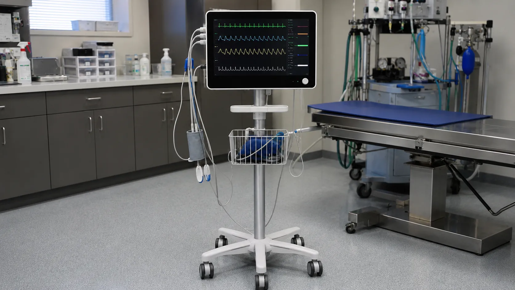
For buyers, depth monitoring comes as standalone units or as modules that plug into an existing anaesthesia monitor. The recurring cost is the single-use forehead sensor, so model consumption against your anaesthetic caseload. Check compatibility with your monitoring platform and anaesthetic machines, the availability of the specific sensors, and that staff are trained to interpret trends rather than chase a single number.
-
Decide whether a standalone monitor or a module for your existing platform fits best.
-
Confirm compatibility with your anaesthesia monitors and machines.
-
Model single-use sensor cost against realistic anaesthetic caseload.
-
Confirm the depth-of-anaesthesia monitor carries UKCA or CE marking and ships with its instructions for use.
-
Verify sensor availability and shelf life for continuity of supply.
-
Ensure staff training covers interpreting trends alongside clinical signs.
-
Define local policy on when depth monitoring is indicated.
Conclusion
Depth of anaesthesia is inferred, not directly read: clinical signs, end-tidal agent against MAC, processed-EEG indices and evoked potentials each give part of the picture, and each has limits. Buying well means choosing monitoring that integrates with your existing kit, budgeting for single-use sensors and training staff to read trends in context. To source anaesthesia monitoring and compatible sensors, register as a buyer or speak to the MediGear team.
Disclaimer
This article is for informational purposes only. It is published by MediGear (medigear.uk) for general information and procurement guidance, and is not clinical, diagnostic, treatment, technical, engineering, legal or regulatory advice, nor a product endorsement, guarantee or substitute for professional assessment. MediGear does not provide medical consultations. Buyers should consult their clinical, biomedical, estates and regulatory contacts, and the manufacturer's documentation, and independently verify all specifications, certifications, compatibility and suitability before purchase. Specifications, certifications and availability are correct at the time of publication and may change without notice. MediGear is a medical-equipment distributor and does not sell medicines or pharmaceutical products.