Carotid intima-media thickness testing uses ultrasound to measure the combined thickness of the two innermost layers of the carotid artery wall, in millimetres, as a marker of cardiovascular risk. Rather than looking for a blockage, a CIMT study measures a wall — a thickened intima-media is an early sign of atherosclerosis, and the number is used to help stratify a patient's long-term cardiovascular risk.
For a buyer, the distinguishing feature is that carotid intima-media thickness is a precise, sub-millimetre measurement, and its accuracy depends far more on the analysis software and technique than on raw imaging power. What you are really procuring is a high-frequency linear probe plus validated automated edge-detection software that can measure a wall to a repeatable fraction of a millimetre.
Why the wall, not the lumen, is the target
A standard carotid duplex scan looks for narrowing of the lumen from plaque. Carotid intima-media thickness testing asks an earlier question: before there is a discrete plaque, is the artery wall already thickening? The intima and media together form a characteristic double-line pattern on a good B-mode image of the far wall of the common carotid, and the distance between those lines is the measurement. Because a thickened wall predates overt disease, CIMT is used as a risk-refinement tool in people who sit in an intermediate risk band, not as a test for stroke-causing stenosis.
How the measurement is actually taken
The technique. The far wall of the common carotid, just below the bulb, is imaged with a high-frequency linear probe at a standardised angle, and the measurement is taken over a defined segment — usually a centimetre — at a fixed point in the cardiac cycle, often gated to the R-wave of a synchronised ECG. Measuring by hand with callipers is slow and operator-dependent, which is why automated edge-detection software has become the norm: it identifies the lumen-intima and media-adventitia boundaries across hundreds of points and reports a mean and maximum thickness with far better reproducibility than a manual read.
The automated edge-detection software is the product.
This is the point that shapes the purchase. Because carotid intima-media thickness values are compared against normal ranges and tracked over time, the measuring software must be validated, consistent, and reproducible. Automated border-detection packages reduce operator variability and produce the mean-thickness and percentile outputs clinicians use. Crucially, values are not perfectly interchangeable between different software algorithms, so a patient followed over time should be measured with the same package. Treat the CIMT software as a regulated medical device in its own right and confirm its validation, version, and reference ranges — not just the scanner it runs on.
The transducer and image quality it needs
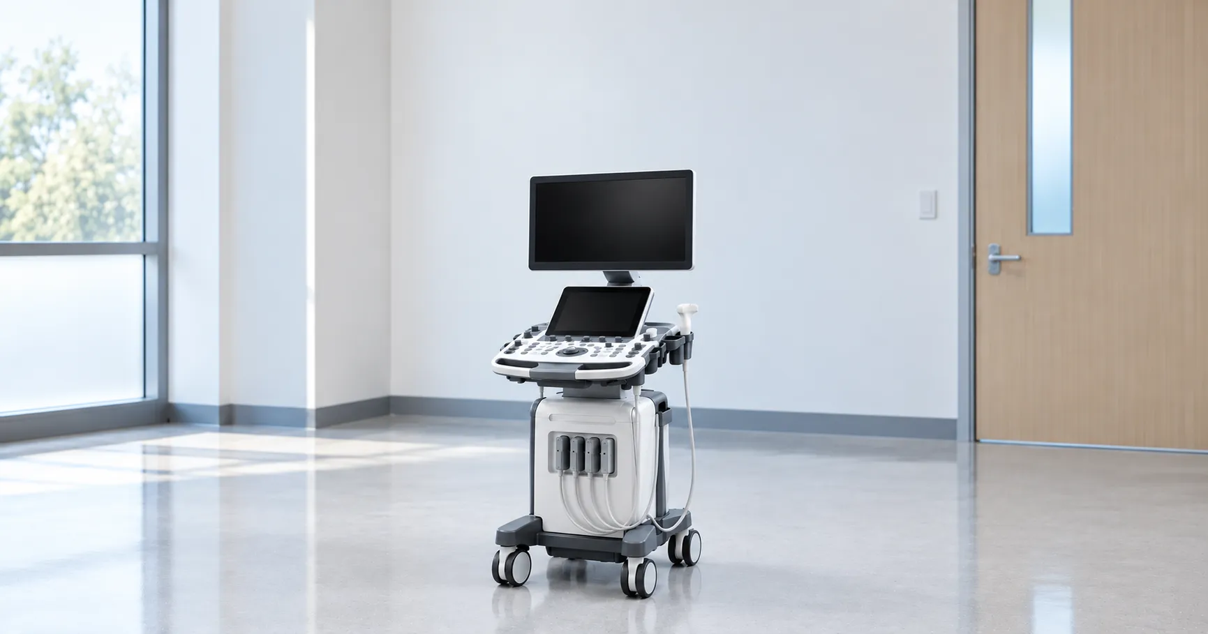
CIMT lives on far-wall image clarity, so a high-frequency linear-array transducer, broadly in the 7–12 MHz range, is essential to resolve the thin double-line at the sub-millimetre scale. The probe must sit on a scanner capable of the frame rate and greyscale detail the edge-detection algorithm needs, and ECG gating helps freeze the wall at a consistent phase. A machine that produces a soft or noisy far-wall image will defeat even good software, so when you trial a system, judge it on a clear common-carotid far wall rather than a glossy demo image.
Where CIMT testing fits in a service
Carotid intima-media thickness testing sits in cardiovascular risk assessment rather than acute vascular diagnosis. Preventive cardiology clinics, lipid and health-screening services and research settings use it to refine risk in intermediate-risk individuals and to track the effect of treatment over years. It does not replace standard carotid duplex for suspected stenosis, and clinical use should follow recognised cardiovascular risk guidance such as that from the National Institute for Health and Care Excellence at nice.org.uk. The procurement decision therefore hinges on whether your service does structured, repeatable risk assessment where a reproducible wall measurement adds value.
Standards, marking and the software question
The scanner is diagnostic ultrasound equipment built to the general standard IEC 60601-1 with the ultrasonic particular standard IEC 60601-2-37 for acoustic output and safety, and it must carry UKCA or CE marking. The carotid intima-media thickness analysis software, because it informs a clinical risk decision, is itself regulated as a medical device and must be UKCA or CE marked. For CIMT hardware and its analysis software, device regulation and field-safety alerts are handled by the Medicines and Healthcare products Regulatory Agency at mhra.gov.uk. This guide draws on manufacturer documentation, UK clinical guidance and the relevant IEC standards, and is written for UK procurement teams.
Reproducibility, protocol and training
A CIMT number is only meaningful if it can be repeated. Reproducibility depends on a fixed protocol — the same wall, segment, angle and cardiac phase every time — and on operators applying it consistently. A service introducing carotid intima-media thickness testing should plan supervised training, a standard operating protocol and a period of double-reading to bed the technique in. Because serial CIMT tracks small changes over years, a small measurement error can swamp a real trend, so consistent acquisition and the same software version matter more here than in most ultrasound work.
Reporting, records and connectivity
The output of a carotid intima-media thickness study is a set of numbers and a percentile, so how it lands in the record matters. Confirm the software exports the mean and maximum thickness, the percentile against its reference population, and the annotated images into your reporting system and PACS. For a screening or preventive service running serial studies, the ability to store and trend results over time is central, not optional, so check the export formats and how prior results are recalled for comparison before you commit.
Beyond the purchase price
Costing CIMT means separating hardware from software. The scanner and probe are the capital item; the analysis package is often a separate, sometimes licensed or subscription, cost with its own updates and validation path. Budget for probe replacement, the service contract, software licensing and upgrades, coupling gel, and the training time that reproducible measurement demands. When you compare a bundled scanner-plus-software offer against adding software to an existing machine, weigh whole-life cost, and use MediGear to compare suppliers on equal terms.
Mean, maximum and where plaque fits in
A carotid intima-media thickness report is usually more than one figure. The mean thickness over the measured segment is the primary risk marker, but the maximum thickness and any focal thickening also matter, and software will often flag a discrete plaque separately once the wall crosses a defined threshold. Some protocols measure only the far wall of the common carotid; others average several angles or include the bulb and internal carotid for a fuller picture. Decide which protocol your clinicians will follow before you buy, because the software must support that measurement scheme — single-wall or multi-angle — and report it against the matching reference population, or the numbers cannot be interpreted.
Pitfalls that distort a CIMT reading
Sub-millimetre measurement is unforgiving of sloppy technique. An off-axis probe angle, a poorly frozen frame, gain set too high or too low, or measuring across a segment that includes a small plaque all shift the number enough to matter. Movement and an ungated frame add error at the fraction-of-a-millimetre scale the test works at. Automated edge-detection reduces the operator's tracing variability but cannot rescue a poor image or a wrongly placed measurement box, so it still needs a trained eye to check every result. When you assess a system, ask how the software behaves on an imperfect far wall, because that is where a CIMT measurement is really won or lost.
Specifying CIMT equipment: buyer's checklist
-
A high-frequency linear transducer, broadly 7–12 MHz, capable of a clear far-wall double-line image.
-
Validated automated edge-detection software with published reproducibility and reference ranges.
-
ECG gating to standardise the cardiac phase of each measurement.
-
UKCA or CE marking for both the scanner and the analysis software, with documentation supplied.
-
Export of mean and maximum thickness, percentile and ima,ges into your reporting system and PACS.
-
A clear software version and update path so serial measurements stay comparable.
-
Training and a standard protocol costed alongside the equipment.
Choosing well for CIMT
Carotid intima-media thickness testing is a precise wall measurement used to refine cardiovascular risk, and its value stands or falls on validated software, a capable high-frequency probe, and a disciplined protocol — not on raw scanner power. Buy the software and the technique as carefully as the hardware, and keep serial measurements on one consistent package. If you are equipping a preventive or screening service, MediGear can help you compare verified suppliers and CIMT software options side by side.
Disclaimer
This article is for informational purposes only. It is published by MediGear (medigear.uk) for general information and procurement guidance, and is not clinical, diagnostic, treatment, technical, engineering, legal or regulatory advice, nor a product endorsement, guarantee or substitute for professional assessment. MediGear does not provide medical consultations. Buyers should consult their clinical, biomedical, estates and regulatory contacts, and the manufacturer's documentation, and independently verify all specifications, certifications, compatibility and suitability before purchase. Specifications, certifications and availability are correct at the time of publication and may change without notice. MediGear is a medical-equipment distributor and does not sell medicines or pharmaceutical products.