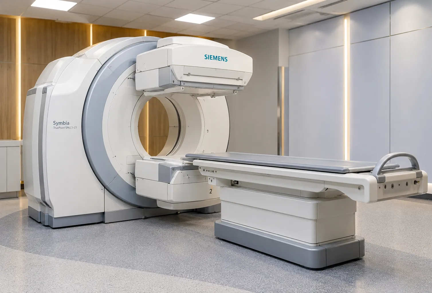
SPECT imaging — single-photon emission computed tomography — turns a series of gamma-camera views taken around the patient into a three-dimensional map of how an injected radiotracer distributes in the body, and in cardiology, that map shows blood flow through the heart muscle. It is the imaging method, not the camera hardware: SPECT is what happens when angular projections are reconstructed into cross-sectional slices and read as a perfusion study. Understanding the technique is what lets a service design protocols that are diagnostic and dose-aware.
In myocardial perfusion imaging, SPECT reveals areas of muscle that receive less blood under stress than at rest — the signature of significant coronary disease. The strength of the method is that it measures function, showing not just where an artery is narrowed but whether that narrowing actually starves the muscle.
From projections to a 3D map: how SPECT reconstruction works
SPECT imaging starts with the detector heads rotating around the patient, collecting many two-dimensional projections at successive angles. A reconstruction algorithm then computes the three-dimensional distribution of tracer that best explains those views. Modern services use iterative reconstruction, typically ordered-subset expectation maximisation, which models the physics of the acquisition and handles noise better than the older filtered back-projection. The reconstructed volume is reoriented along the heart's own axes into short-axis, vertical and horizontal long-axis slices, the standard views a reporter reads. Reconstruction settings materially affect image quality, so they are part of protocol governance, not an afterthought. Resolution-recovery and scatter-correction options built into modern iterative algorithms can recover count statistics, which is what lets high-sensitivity services shorten acquisition times or cut tracer dose without losing diagnostic confidence.
Myocardial perfusion imaging: stress and rest
A perfusion study compares the heart under two states. Stress imaging, achieved by exercise or a pharmacological agent that dilates the coronary arteries, is paired with a rest study, and the two SPECT datasets are compared. A defect present on stress but not at rest suggests reversible ischaemia; a defect fixed on both may indicate scar. Protocols can be one-day or two-day, and can image rest or stress first, each with trade-offs in tracer dose, patient time and department flow. Designing the protocol around your caseload is central to running an efficient service.
Tracers that make perfusion visible
SPECT imaging depends on the radiopharmaceutical injected. Technetium-99m agents — sestamibi and tetrofosmin — are the mainstay for cardiac perfusion, offering good imaging characteristics and a short half-life that keeps patient dose reasonable. Thallium-201 is an older tracer still used in some viability work. Tracer choice interacts with protocol design, camera sensitivity and dose, and every administration must be justified and authorised. Because these are unsealed radioactive substances, their supply, storage and use sit within strict radiation governance rather than ordinary pharmacy stock. Technetium is typically eluted from a generator on site, so reliable supply logistics and a compliant hot lab are as much a part of running SPECT imaging as the camera itself.
Attenuation correction and artefact control
Soft tissue absorbs gamma photons on their way to the detector, and this attenuation can mimic a perfusion defect — the diaphragm or breast tissue casting an artefactual shadow. Managing it is part of good SPECT imaging. CT-based attenuation correction, available on hybrid SPECT/CT systems, maps tissue density and corrects the data. Where that is not available, prone imaging and careful interpretation help distinguish artefact from real disease. Recognising and correcting attenuation is one of the biggest determinants of diagnostic accuracy in myocardial perfusion.
Gated SPECT: adding function to perfusion
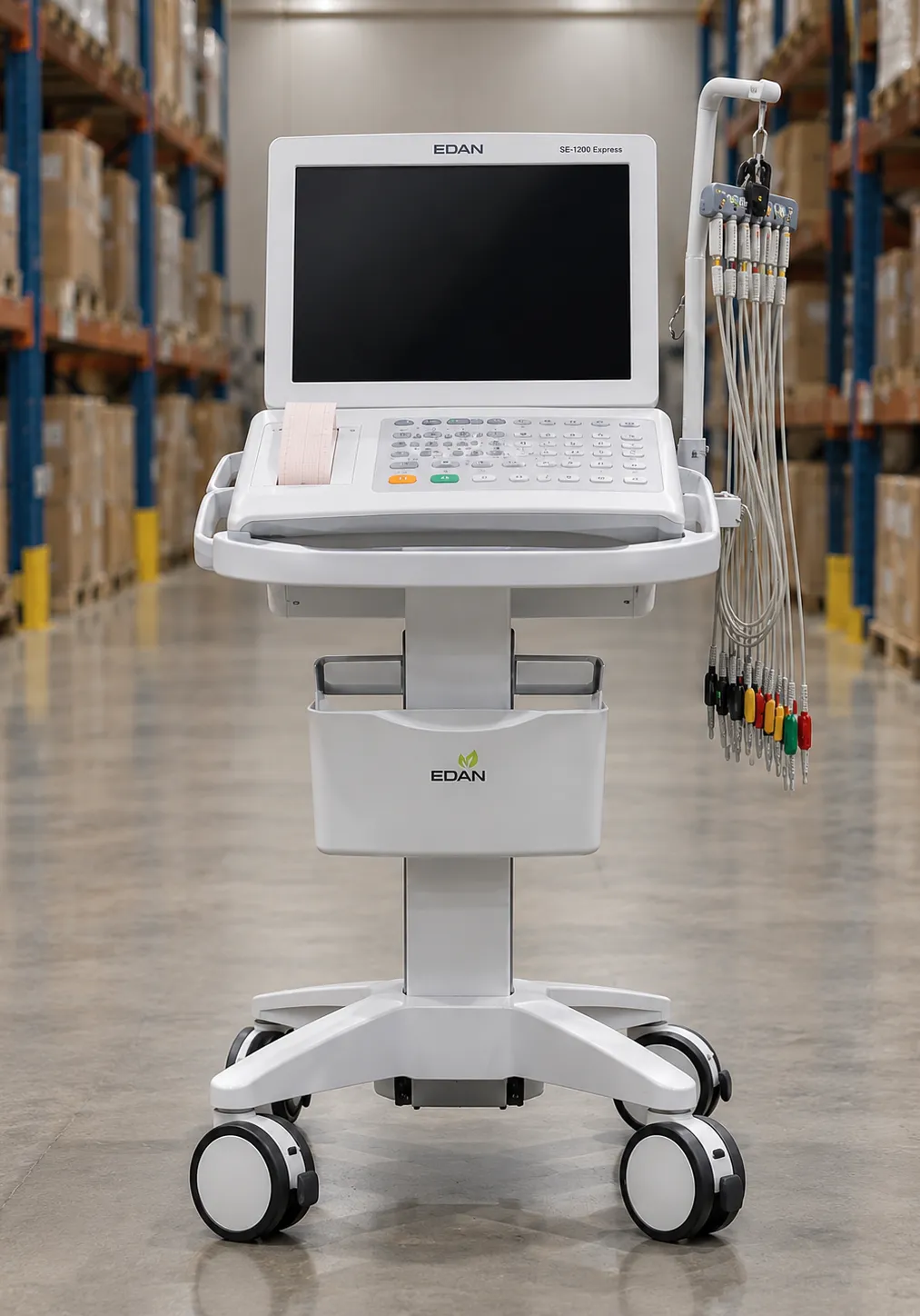
Synchronising acquisition to the ECG lets SPECT imaging report on function as well as flow. Gated SPECT divides the cardiac cycle into frames, so the same study yields left-ventricular ejection fraction, wall motion and wall thickening alongside the perfusion pattern. That combined read strengthens the diagnosis and adds prognostic information without a second test — one reason gated acquisition is now routine in myocardial perfusion imaging.
Where SPECT imaging fits in the cardiac pathway
SPECT myocardial perfusion imaging is a well-established test for known or suspected coronary artery disease, used to assess the functional significance of stenoses and to guide whether a patient needs revascularisation. Its place among competing tests — CT coronary angiography, stress echocardiography and cardiac MRI — is set out in the clinical guidance from the National Institute for Health and Care Excellence at nice.org.uk. Aligning your service to those pathways underpins the business case, and MediGear can help you shortlist verified suppliers once the clinical need is clear.
The regulatory frame around a SPECT service
Every SPECT imaging study exposes a patient to ionising radiation from the tracer, so the method is tightly governed. The Ionising Radiation (Medical Exposure) Regulations 2017 require each exposure to be justified and optimised, and administering the radiopharmaceutical needs an ARSAC licence — both covered in UK government guidance at gov.uk. Staff protection and unsealed-source handling fall under the Ionising Radiations Regulations 2017, overseen by the Health and Safety Executive at hse.gov.uk. Protocols, dose reference levels and quality control are therefore core to commissioning, not optional extras.
Stress agents: exercise versus pharmacological
The stress half of a perfusion study can be produced two ways. Exercise on a treadmill or bike raises cardiac demand physiologically and adds information about symptoms and capacity. Where a patient cannot exercise adequately, a pharmacological agent is used instead: a vasodilator such as adenosine, regadenoso,n or dipyridamole widens the coronary arteries so that diseased territories fail to increase flow, or dobutamine to raise demand directly. The tracer is injected at peak stress and the SPECT images acquired shortly after. Choice of agent affects patient monitoring, contraindications and workflow, and it belongs in protocol design rather than being left to the day.
Quantification, polar maps and reporting
Modern SPECT imaging does not stop at pictures. Software segments the left ventricle and expresses tracer uptake as a polar map, or bull's-eye, comparing each region against a normal database and putting a number on the size and severity of a defect. Quantitative perfusion and function scores sharpen consistency between reporters and support serial comparison over time. When commissioning a service, weigh the reporting package and its normal databases as heavily as the acquisition, because that is where much of the diagnostic value is realised.
Protocol design and dose optimisation
How a service configures SPECT imaging drives both quality and patient dose. One-day and two-day protocols, stress-first strategies, weight-adjusted tracer activity and the faster acquisitions that high-sensitivity cameras allow all shift the balance between throughput, image quality and exposure. The regulations require exposures to be optimised, so a good service audits its dose against diagnostic reference levels and reviews protocols regularly. Reconstruction settings, acquisition time and tracer activity are levers to be tuned together, not fixed once and forgotten.
What buyers should verify before commissioning SPECT imaging
-
Reconstruction software — iterative/OSEM with resolution recovery and noise handling for cardiac work.
-
Attenuation-correction route, whether CT-based hybrid correction or a validated prone protocol.
-
Gated SPECT capability for ejection fraction and wall-motion analysis in the same study.
-
Stress and rest protocol options, one-day and two-day, matched to your throughput and dose targets.
-
Tracer supply, storage and ARSAC-authorised administration arrangements.
-
Quality-control and dose-audit tools to satisfy IR(ME)R 2017 and IRR 2017.
-
Reporting workstation, reorientation tools and PACS integration for slice review.
-
Training and applications support so staff can run and interpret perfusion studies reliably.
Where SPECT imaging can fall short
No test is perfect, and knowing the limits of SPECT imaging protects a service from over-reading. Balanced multi-vessel disease can look deceptively uniform because there is no normal territory to compare against, and attenuation artefact still fools the unwary reader. Spatial resolution is coarser than cardiac MRI or CT, and patient motion during the rotation degrades the reconstruction. These are reasons to invest in attenuation correction, gating and staff training, and to keep the test within the pathways where its function-first strength genuinely helps the decision.
The verdict for perfusion services
SPECT imaging remains a dependable, function-first way to test the heart for ischaemia, and its value rests on the method rather than the box: sound reconstruction, honest attenuation correction, gated acquisition and well-designed stress and rest protocols. This guide draws on manufacturer documentation and UK regulatory guidance and is written for procurement and clinical-engineering teams. If you are scoping a perfusion service, MediGear can help you compare verified suppliers on the specifications that decide diagnostic quality.
Disclaimer
This article is for informational purposes only. It is published by MediGear (medigear.uk) for general information and procurement guidance, and is not clinical, diagnostic, treatment, technical, engineering, legal or regulatory advice, nor a product endorsement, guarantee or substitute for professional assessment. MediGear does not provide medical consultations. Buyers should consult their clinical, biomedical, estates and regulatory contacts, and the manufacturer's documentation, and independently verify all specifications, certifications, compatibility and suitability before purchase. Specifications, certifications and availability are correct at the time of publication and may change without notice. MediGear is a medical-equipment distributor and does not sell medicines or pharmaceutical products.