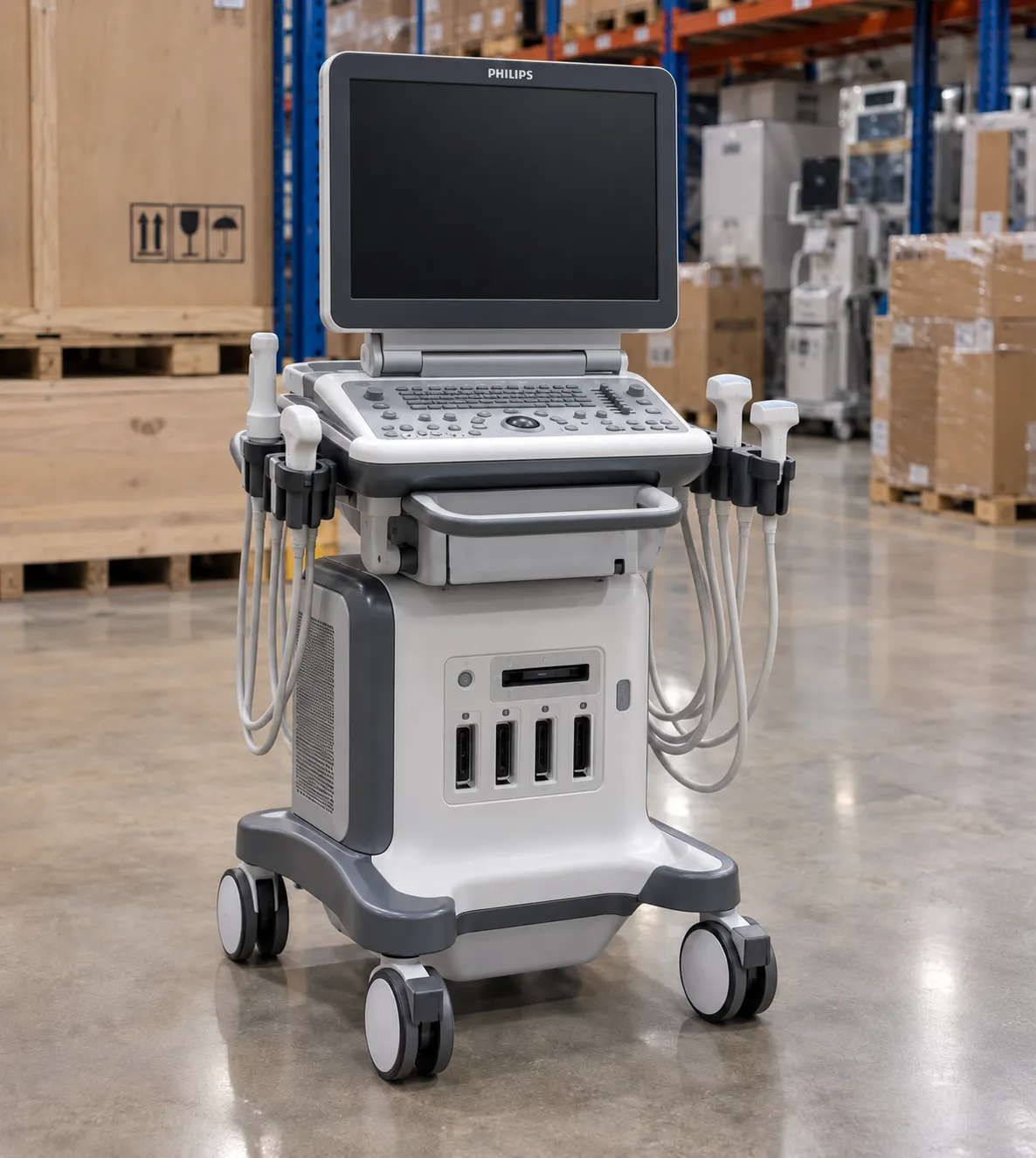
An echocardiography machine is a cardiac ultrasound system that turns high-frequency sound into live images of the beating heart, letting clinicians measure chamber size, watch the valves move, and judge how strongly the muscle pumps. Radiation-free, portable,e and quick to set up, it is the workhorse of cardiology and acute care, and for most patients it is the first heart scan performed — a transthoracic study built from a phased-array probe, greyscale 2D imaging, and a full Doppler suite.
For a procurement lead,d the echocardiography machine is really a bundle of decisions: the console and its image processing, the transducers, and the licensed measurement packages. Get the transducer set and Doppler modes right and one system covers a full transthoracic list. Skimp on them and you have bought an expensive picture box that cannot report a study properly.
How a transthoracic echo builds its picture
Standard scanning is transthoracic — the probe sits on the chest wall and images through and between the ribs. A phased-array transducer steers a sound beam electronically across a wedge-shaped sector, and the machine times the returning echoes to build a moving two-dimensional image many times a second. M-mode takes a single line through that image and plots it against time, giving the precise timing and dimensions clinicians still use for wall thickness and chamber measurement. Tissue harmonic imaging, now standard, listens at twice the transmitted frequency to cut clutter and sharpen the endocardial border, which matters on difficult chests. Where that border is still unclear, contrast echocardiography — a licensed option that opacifies the chamber with an intravenous microbubble agent — defines the endocardium and firms up ejection-fraction and wall-motion reads. Confirm whether a system supports contrast imaging if your service scans many difficult chests.
Doppler: turning echoes into blood-flow data
An echocardiography machine is not just anatomy; the Doppler modes make it a haemodynamic tool. Colour Doppler overlays flow direction and turbulence on the 2D image, so a leaking valve or a shunt shows as a coloured jet. Pulsed-wave Doppler samples velocity at a chosen depth for gentle flows; continuous-wave Doppler measures the high velocities across a narrowed valve without aliasing. Tissue Doppler applies the same principle to the moving muscle to assess diastolic function. A machine sold without the full Doppler suite cannot complete a standard adult study, so confirm every mode is licensed, not merely fitted.
Transducers decide what the machine can scan.
The probe is where image quality is won or lost. A phased-array cardiac transducer, typically in the 1–5 MHz range, is the core; lower frequencies penetrate deeper for large patients, higher frequencies sharpen near-field detail in children. A paediatric service needs a higher-frequency probe, and many departments add a small-footprint probe for portable work. Some consoles also drive a transoesophageal echo probe for the semi-invasive views a chest-wall scan cannot reach — a separate purchase with its own reprocessing burden covered in its own guide. Count the probe ports and confirm the transducers you need are included, because probes are among the most expensive and most easily damaged parts of the system.
3D, matrix probes and quantification
A 3D matrix-array transducer carries thousands of elements and can acquire a pyramid of data for real-time three- and four-dimensional echo — live 3D of a moving valve — valuable for structural and valve assessment and for accurate, geometry-independent chamber volumes. Modern machines add automated quantification packages: border detection that measures ejection fraction, automated chamber quantification, and speckle-tracking strain to gauge subtle changes in wall motion the eye misses. Those packages are usually licensed options. Decide which your reporting clinicians actually use before paying for a full analytics suite, and check whether the licence is perpetual or subscription.
Cart-based, portable and handheld formats
Echocardiography machines span a wide range of formats. A full cart-based console gives the best image processing, the widest probe support and the deepest measurement packages for a dedicated echo lab. Compact and laptop-style systems trade some processing for portability on wards and in clinics. Handheld pocket scanners suit focused bedside looks but are not a substitute for a full diagnostic study. Match the format to where the scanning happens; many services run a cart in the lab and lighter systems for ward and theatre cover.
Standards, marking and electrical safety
As medical electrical equipment,t an echocardiography machine is built to the general standard IEC 60601-1, with the particular standard for ultrasonic diagnostic equipment, IEC 60601-2-37, covering acoustic output and safety. The device must carry UKCA or CE marking, and any purchase should come with conformance documentation you can file. Device regulation and field-safety notices sit with the Medicines and Healthcare products Regulatory Agency at mhra.gov.uk, and checking a manufacturer's alert history there is a sensible pre-purchase step. This guide draws on manufacturer specifications, the IEC 60601 family and UK regulatory guidance, and is written for UK procurement and clinical-engineering teams rather than patients.
Probe care and everyday hygiene
Transthoracic probes touch intact skin, so they need low-level cleaning and disinfection between patients following the manufacturer's compatible-agent list — harsh wipes crack the lens and void the warranty. That is a far lighter burden than a semi-invasive probe, but it still needs a documented protocol and staff who follow it. Confirm the manufacturer's approved disinfectant list before standardising your wipes, because using the wrong chemical is a common cause of premature transducer failure.
Connectivity, DICOM and reporting workflow
An echo study is only useful once it reaches the record. Confirm the machine exports DICOM cleanly to your PACS and cardiology information system, supports your structured reporting, and handles worklists so studies attach to the right patient automatically. Wired or wireless transfer, storage capacity, and the reporting package all shape how quickly a sonographer turns a list around. Integration gaps are a common, avoidable drag on a busy department.
Beyond the purchase price
On an echocardiography machine, the console is only the opening figure in the budget. Transducers fail, get dropped,d and need replacing; a single cardiac probe is a significant sum, so weigh probe warranty and replacement terms heavily. Budget for the service contract, software upgrades, cleaning consumables, and the eventual battery replacement on portable units. When you compare quotes, compare whole-life cost and use MediGear's tools to weigh suppliers on equal terms rather than headline price.
Depth, frame rate and image optimisation
Two patients are never the same, so an echocardiography machine lives or dies on its optimisation controls. Depth, gain, sector width, focus and dynamic range let the operator tune the image to the chest in front of them, while a higher frame rate freezes a fast heart and a narrower sector buys frame rate back on a tachycardic patient. Good machines make these adjustments quick and offer sensible presets for adult, paediatric and difficult studies. When you trial a system, scan a genuinely tricky patient rather than an easy volunteer, because the gap between a basic and a capable machine shows most on the hard chests that make up much of a real list.
Ergonomics and the sonographer's working day
Echocardiography is repetitive, physically demanding work, and musculoskeletal injury among sonographers is a real and costly problem for a service. The machine's ergonomics matter more than most buyers expect: an adjustable control panel and monitor arm, a light and well-balanced transducer, and a low-effort keyboard all reduce the strain of a full scanning list. Sit-stand consoles and configurable examination presets that cut keystrokes help a department protect its staff and keep throughput up. Trial the machine with the sonographers who will use it daily, not just the reporting clinicians, because a system that scans awkwardly will slow every list and shorten careers.
New and refurbished echocardiography machines
Not every echocardiography machine is bought new. A well-supported refurbished console from a reputable supplier can stretch a capital budget, provided it comes with a warranty, current software and a documented service history. The more important question for any purchase, new or used, is the upgrade path: can the platform take future software licences and newer transducers, and how long will the manufacturer support it with spares and security updates? A machine that cannot be upgraded ages quickly. Confirm the end-of-support date and the cost of the licences you are likely to want before you commit, so the platform still fits your service in five years.
Specifying an echocardiography machine: buyer's checklist
-
Phased-array cardiac transducer range matched to your patients, plus paediatric or portable probes if needed.
-
Full Doppler suite — colour, pulsed-wave, continuous-wave and tissue Doppler — licensed, not just fitted.
-
Tissue harmonic imaging and the measurement packages your reporters actually use.
-
3D or matrix capability and automated quantification only if your service will use them.
-
Number of active probe ports and which transducers are included in the price.
-
IEC 60601-2-37 compliance on the cardiac scanner, with valid UKCA or CE marking and the conformance paperwork handed over.
-
DICOM export, PACS and cardiology-system integration, and structured reporting support.
-
Probe warranty and replacement terms, service cover and a clear upgrade path.
Choosing well for your echo service
An echocardiography machine is a long-term clinical workhorse, so judge it on the whole picture: the transducers and Doppler modes that let it complete a full study, the software your reporters will genuinely use, and the probe and service costs that follow the invoice. Specify it properly, ly and one system will carry the bulk of your heart-imaging work for years. If you are scoping a machine, MediGear can help you reach the right suppliers and compare them fairly.
Disclaimer
This article is for informational purposes only. It is published by MediGear (medigear.uk) for general information and procurement guidance, and is not clinical, diagnostic, treatment, technical, engineering, legal or regulatory advice, nor a product endorsement, guarantee or substitute for professional assessment. MediGear does not provide medical consultations. Buyers should consult their clinical, biomedical, estates and regulatory contacts, and the manufacturer's documentation, and independently verify all specifications, certifications, compatibility and suitability before purchase. Specifications, certifications and availability are correct at the time of publication and may change without notice. MediGear is a medical-equipment distributor and does not sell medicines or pharmaceutical products.