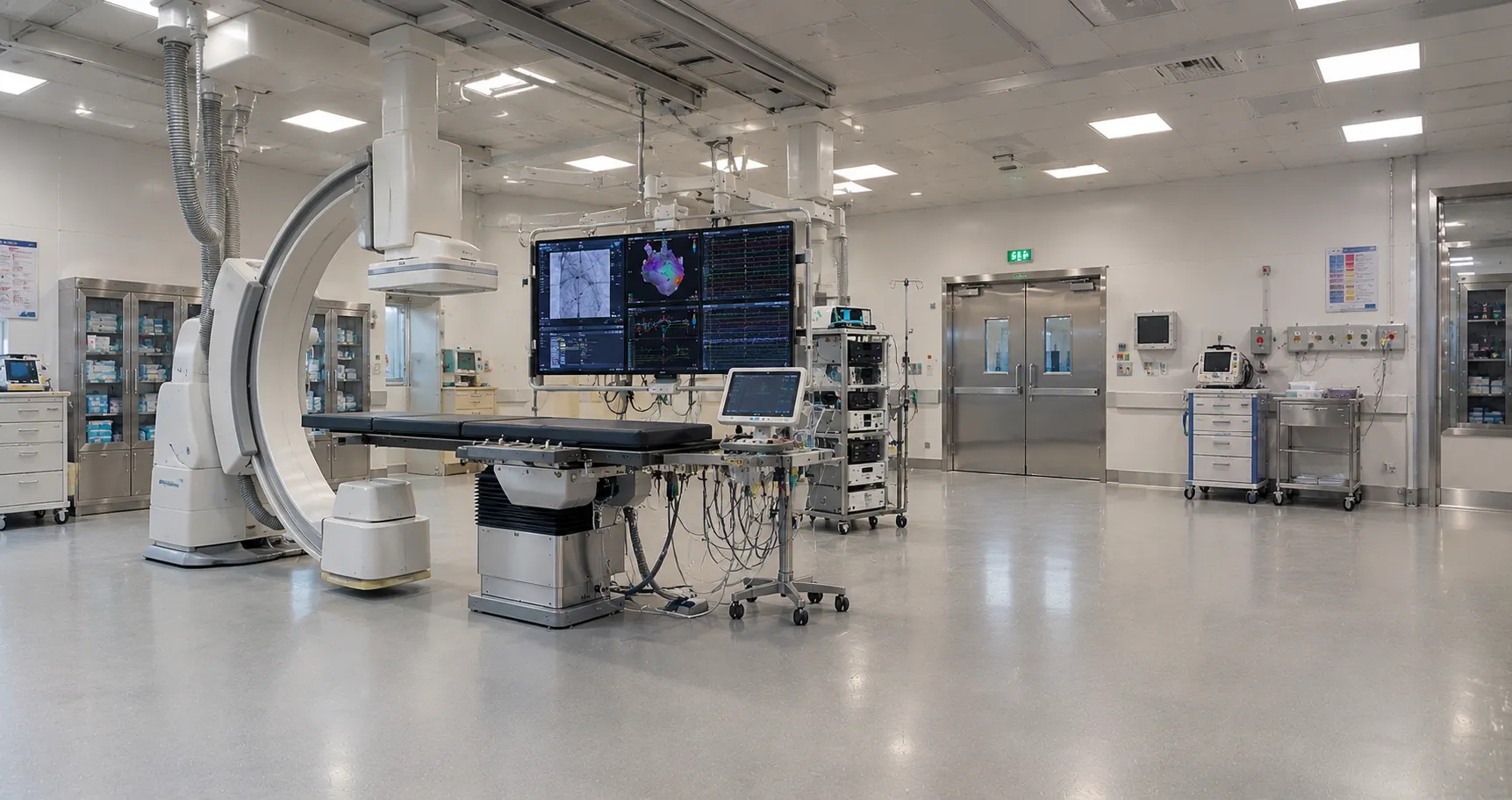
The single specification that defines a modern electrophysiology system is mapping resolution: how precisely it can build a three-dimensional model of a heart chamber and pinpoint where an abnormal rhythm begins. Electrophysiology systems are recording, mapping, and ablation platforms used to diagnose arrhythmias and then treat them by burning or freezing tissue. They share a room with the general cath lab and its fluoroscopy, but the defining kit here is different: a multichannel EP recorder, a 3D electroanatomical mapping system and an energy generator that delivers the ablation. Get the mapping and ablation chain right, and fluoroscopy becomes a supporting role rather than the main tool.
This article foregrounds mapping and ablation. General diagnostic angiography and intracoronary ultrasound are separate equipment covered elsewhere, so the focus stays on the arrhythmia workflow.
The EP recording system and its signals
At the base of every electrophysiology system is a multichannel recorder that captures intracardiac electrograms alongside the surface ECG at high sampling rates. During a study, catheters placed at defined sites in the heart feed dozens of channels into this recorder, and the electrophysiologist reads the timing between signals to work out the arrhythmia mechanism. Signal fidelity, channel count, and low-noise amplification matter, because the diagnosis rests on millisecond differences between electrograms. A programmed stimulator is part of the same platform, delivering precisely timed pacing to provoke and characterise the rhythm under study.
Three-dimensional mapping that guides the ablation
The feature that transformed electrophysiology systems is 3D electroanatomical mapping. Rather than relying on X-ray alone, the mapping system tracks catheter position in space, using magnetic fields or impedance sensing, and paints a co-created model of the chamber showing the electrical activation sequence and scar areas, letting the operator see exactly where to ablate and to return to the same spot, which cuts fluoroscopy time and dose for both patient and staff. Mapping accuracy, catheter compatibility and how well the system handles complex arrhythmias such as atrial fibrillation are the real differentiators between platforms, and they should drive the choice far more than headline price.
Ablation energy: radiofrequency, cryo and beyond
Treatment is Anator paired with a catheter that delivers treatment through ablation. Radiofrequency ablation heats tissue through a catheter tip to create a controlled lesion and is the long-established workhorse; cryoablation freezes tissue, often via a balloon catheter used to isolate the pulmonary veins in atrial fibrillation. Newer pulsed-field ablation uses electrical pulses designed to target cardiac tissue selectively. Each modality needs its own generator and catheters, so the energy strategy shapes both the capital purchase and the recurring consumable bill. Match the generator to the arrhythmias your service treats rather than buying capability you will not use.
Radiation, shielding and the shared fluoroscopy
Even a mapping-heavy electrophysiology procedure still uses fluoroscopy, so radiation protection remains part of the specification. The exposure sits under the Ionising Radiations Regulations 2017 for staff and the Ionising Radiation (Medical Exposure) Regulations 2017 for patients, both set out in the guidance on GOV.UK. Good 3D mapping reduces reliance on X-ray, but ceiling-suspended lead screens, dose monitoring and recorded dose-area product still belong in the room. Confirm how the mapping system, recorder and imaging chain integrate, because disjointed kit costs time in long procedures and time under fluoroscopy is dose.
Comparing mapping platforms before you buy
Choosing between electrophysiology systems is mostly a choice between mapping platforms, and the demonstration case rarely tells the whole story. Look at how the system builds a map from a walk-around versus a multipolar catheter, how quickly it acquires points, and how it copes when the rhythm is unstable, as in persistent atrial fibrillation. High-density mapping catheters capture thousands of points fast but add cost quickly, so weigh the clinical benefit against overall price and your case mix. Compatibility matters too: some mapping systems work with a wide range of third-party catheters, others are tightly closed, and that openness affects both flexibility and long-term cost. Insist on a hands-on evaluation with your own operators and typical cases rather than relying on a curated vendor demonstration.
Training, software and building the service
An electrophysiology system is only as good as the team running it, so training and software support are part of the purchase, not an afterthought. Mapping platforms have a learning curve, and vendors differ in how much proctoring, application, and ongoing support they provide as staff and software evolve. Software is central here: mapping algorithms and analysis tools are updated, and the upgrade path, licensing model, and cost of future modules can shape the platform's usefulness for years. Confirm what training is included at installation, how updates are delivered and priced, and what applications cover you can call on during complex cases. Building an EP service is a multi-year commitment, and the supplier relationship matters as much as the hardware on day one.
Standards and regulation for electrophysiology systems
An electrophysiology system is medical electrical equipment built to the general standard IEC 60601-1, with the fluoroscopy meeting IEC 60601-2-43 for interventional X-ray and the ablation generator treated as high-frequency surgical equipment under its associated particular standards. All components must carry UKCA or CE marking and are regulated under the UK Medical Devices Regulations 2002, overseen by the device regulator, the MHRA. This guide is drawn from manufacturer documentation and UK regulatory sources and is written for procurement teams; the clinical selection of mapping and ablation strategy rests with the electrophysiology team.
Data integration and reporting
An electrophysiology study generates a large volume of signal and mapping data, and how the system stores, reports and shares it affects both efficiency and audit. The EP recorder should archive electrograms and events in a format that can be reviewed later, and the mapping system should export images and measurements into the procedure report and the patient record rather than keeping them isolated. The recorder, mapping platform, and reporting tools should connect, allow remote review for teaching or audit, and support the outcome tracking a growing arrhythmia service needs. Clean data flow reduces manual work after every case and supports the service governance commissioners increasingly expect, so treat integration as a core requirement, not a technical detail.
Specifying an electrophysiology system: buyer checklist
Work through the points that decide whether the platform fits your arrhythmia service:
-
Mapping accuracy and how the system handles complex atrial fibrillation cases.
-
Recorder channel count, sampling rate and electrogram signal quality.
-
Ablation modality support, frequency, cryo or pulsed-field, and matching catheters.
-
Catheter and consumable compatibility, shelf life and resupply reliability.
-
Integration between recorder, mapping system and the room's fluoroscopy.
-
Radiation shielding and recorded dose monitoring for long procedures.
-
IEC 60601-1 and IEC 60601-2-43 conformity plus UKCA or CE marking.
-
Software upgrade path, training provision and engineer response times.
Beyond the capital cost of ablation equipment
Electrophysiology systems carry a substantial recurring cost that dwarfs many disposable lines. Mapping catheters, ablation catheters, cryoballoons, and disposable patches are used in every case, and each mapping platform tends to lock you into its family, shaping your bill for years. Software licensing and periodic upgrades add to the total, as do service contracts on the generator and mapping hardware. When you compare vendors, model the per-case consumable spend alongside the capital and licensing, and line up EP suppliers through MediGear so the whole cost picture is visible before you commit.
The bottom line for buyers
Electrophysiology systems live or die on mapping accuracy and a coherent recording-to-ablation workflow, with fluoroscopy as backup rather than the primary guide. Match the mapping and energy platform to the arrhythmias you treat, weigh the locked-in consumable cost, and confirm the standards before purchase. As a UK medical-equipment distributor, MediGear helps facilities compare compliant EP platforms and their catheter supply; begin a specification with us via the MediGear enquiry desk.
Disclaimer
This article is for informational purposes only. It is published by MediGear (medigear.uk) for general information. It is not medical or technical advice, nor is it regulatory advice or a substitute for professional assessment. MediGear does not provide medical consultations. Buyers should consult their clinical, biomedical, estates and regulatory contacts, and the manufacturer's documentation, and independently verify all specifications, certifications, compatibility and suitability before purchase. Specifications, certifications and availability are correct at the time of publication and may change without notice. MediGear is a medical-equipment distributor and does not sell medicines or pharmaceutical products.