Cardiac catheterisation is often pictured as a single machine, but it is really a procedure delivered by a room full of coordinated equipment. Cardiac catheterisation means threading thin catheters through a wrist or groin artery up into the heart and coronary vessels to measure pressures, inject contrast and, when needed, treat a narrowing. The equipment that makes this possible is the cath lab: an imaging C-arm, a floating-top angiography table, a haemodynamic recording system, a contrast injector and the disposable catheters and wires themselves. Understanding those parts is the difference between buying a coherent lab and assembling a room that fights itself.
This piece stays on the general diagnostic and interventional cath lab. The specialised 3D mapping used for arrhythmia ablation and the intracoronary ultrasound used to size vessels are distinct kit, covered separately.
The imaging C-arm at the heart of the cath lab
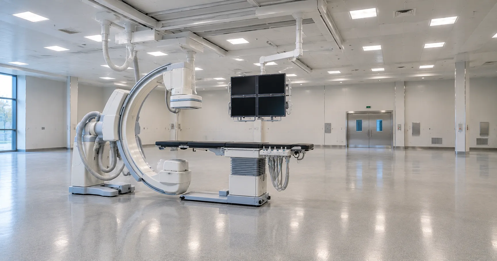
Every cardiac catheterisation depends on live X-ray, so the fixed C-arm angiography system is the defining capital item. A flat-panel detector produces the fluoroscopic images that let the operator steer catheters in real time and the higher-dose cine runs that document coronary anatomy. Detector size, image processing and, above all, dose-reduction technology separate one system from another, because the operator and staff stand in that scatter field for every case. Single-plane systems suit most adult coronary work; biplane rooms, imaging in two directions at once, are specified for complex structural and paediatric cases where contrast and time must be minimised.
Table, haemodynamics and the contrast injector
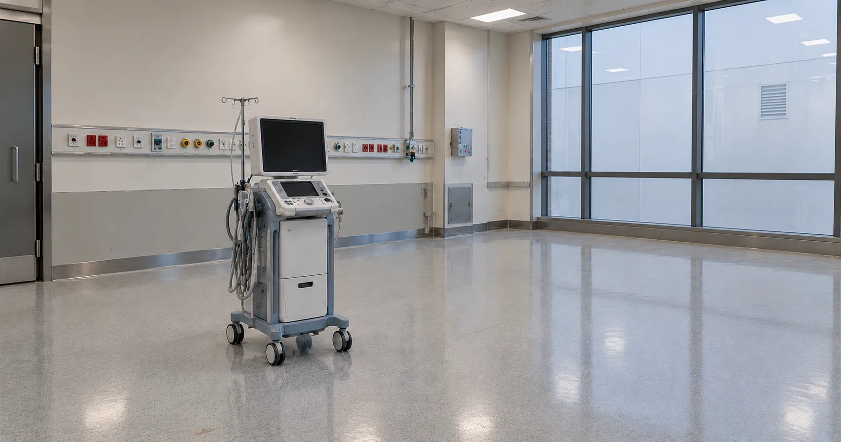
The angiography table is a precision device, not furniture. Its floating, motorised top must move smoothly and lock firmly so the operator can pan across the anatomy without losing the catheter tip, and it has to be radiolucent so the X-ray beam passes cleanly. Alongside it, the haemodynamic monitoring system records intracardiac pressures, ECG and oxygen saturations, turning waveforms into the numbers that define a diagnosis. A power contrast injector delivers a controlled bolus of iodinated contrast for cine angiography. These three elements have to talk to each other and to the imaging chain, which is why integration is a procurement priority rather than a bonus.
Catheters, wires and the single-use inventory
The consumable side of cardiac catheterisation is large and clinically critical. Diagnostic and guide catheters come in a range of shapes and French sizes to reach different coronary origins; guidewires, introducer sheaths, manifolds, contrast and haemostasis devices are all single-use. This inventory is a standing cost and a supply-chain risk, so shelf life, compatibility with your imaging and injector, and reliable resupply matter as much as unit price. A lab that runs out of a specific guide shape mid-list is a lab that cancels patients, which is why buyers often compare catheter suppliers through MediGear before committing to a single source.
Radiation protection and dose management
Because cardiac catheterisation uses ionising radiation on staff who are present for every case, radiation safety is central to the equipment specification. In Great Britain the exposure is governed by the Ionising Radiations Regulations 2017 for worker protection and the Ionising Radiation (Medical Exposure) Regulations 2017 for the patient, both explained in the legislation and guidance on GOV.UK. Practically, that means ceiling-suspended lead screens, table-mounted skirts, dose-area-product monitoring and a system that displays and records cumulative dose. Specify the shielding and dose-reporting features as hard requirements, not extras, and confirm your medical physics team is engaged from the outset.
Access, staffing and where cardiac catheterisation runs
Cardiac catheterisation has moved steadily toward radial access through the wrist, which lowers bleeding risk and lets many patients go home the same day, so the lab supports a day-case flow with recovery bays rather than long inpatient stays. That pathway shapes the room around the imaging: haemostasis devices, monitored recovery and clear discharge criteria all feed off the core equipment. Services also vary in scope, from diagnostic-only angiography suites to full interventional labs performing angioplasty and stenting, and increasingly hybrid rooms shared with structural and surgical work. Understanding which of these your service is buying for prevents over- or under-specifying the room, and the wider NHS pathway context for coronary disease is set out on the NHS website rather than in any single equipment brochure.
Installation, room build and physics acceptance
A cath lab is as much a building project as an equipment purchase. The room needs lead-lined walls and doors, adequate power and cooling for the X-ray generator, and a floor able to carry a ceiling-suspended or floor-mounted C-arm and a heavy table. Before the first patient, a critical examination and acceptance testing by medical physics confirms the system performs to specification and that dose is optimised. Factor this build cost, the installation downtime and the physics sign-off into the project timeline, because a system that is cheap to buy but slow and costly to commission can undermine the whole business case. Engage estates and radiation protection colleagues from the earliest scoping stage.
Standards and regulation for a cath lab
The interventional X-ray system is medical electrical equipment built to the general standard IEC 60601-1 and, specifically, to IEC 60601-2-43, the particular standard for X-ray equipment used in interventional procedures. Every device in the room, from the injector to the catheters, must carry UKCA or CE marking and is regulated under the UK Medical Devices Regulations 2002, overseen by the device regulator, the MHRA. This guide is compiled from manufacturer documentation and UK regulatory sources for procurement teams; clinical protocols and dose optimisation remain with the cardiology and medical physics staff who run the lab.
Image quality and the operator's view
Everything in cardiac catheterisation is done to what the operator can see on the monitor, so image quality and the display setup deserve close attention. A good system balances sharp, low-noise images against the lowest practical dose, and large ceiling-mounted displays that combine live fluoroscopy with reference images and haemodynamic traces help the operator work without turning away from the field. Ask how the system processes images to hold detail at reduced dose, how it stores and recalls cine runs, and whether the archive links to your wider imaging network for review and reporting. A room that produces clear images at low dose protects both the patient and the staff who stand in scatter for every case, and it makes complex work faster and safer.
Specifying a cath lab: what to verify
Treat the room as a system when you go to market:
-
Single-plane or biplane imaging, matched to the case mix you plan to run.
-
Dose-reduction technology and the recorded dose-area-product reporting.
-
Table load rating, movement range and true radiolucency of the top.
-
Integration between imaging, haemodynamics and the contrast injector.
-
Ceiling and table radiation shielding specified as a firm requirement.
-
Catheter and guidewire supply: French sizes, shelf life and resupply lead time.
-
IEC 60601-2-43 conformity, UKCA or CE marking and the physics acceptance test.
-
Service cover, X-ray tube warranty and detector replacement cost.
Beyond the purchase price of a cath lab
A cath lab is one of the larger capital purchases a cardiology service makes, and the sticker price hides the real spend. The X-ray tube and flat-panel detector are expensive wear items with finite lifespans, service contracts are substantial, and the single-use catheter inventory recurs with every case. Room build, lead-lined walls and medical physics acceptance testing add to the first-year total. When you compare vendors, weigh tube and detector lifetime, contracted uptime and consumable pricing together, because a modest saving on capital can vanish against a costly service agreement.
The verdict for procurement teams
Cardiac catheterisation is delivered by an integrated lab, and the equipment decision succeeds or fails on how well the imaging, table, haemodynamics, injector and consumables work as one, under proper radiation control. Specify the system, not the components in isolation, and bring medical physics in early. As a UK medical-equipment distributor, MediGear helps facilities compare compliant imaging systems and catheter suppliers; scope a cath-lab requirement with us through the MediGear enquiry desk.
Disclaimer
This article is for informational purposes only. It is published by MediGear (medigear.uk) for general information and procurement guidance, and is not clinical, diagnostic, treatment, technical, engineering, legal or regulatory advice, nor a product endorsement, guarantee or substitute for professional assessment. MediGear does not provide medical consultations. Buyers should consult their clinical, biomedical, estates and regulatory contacts, and the manufacturer's documentation, and independently verify all specifications, certifications, compatibility and suitability before purchase. Specifications, certifications and availability are correct at the time of publication and may change without notice. MediGear is a medical-equipment distributor and does not sell medicines or pharmaceutical products.