Veterinary X-ray in a small animal practice has its own character, shaped by patients who must be positioned and held still and by a wide range of body sizes. A unit suited to veterinary work is one whose detector, positioning, and image-handling capabilities fit that setting, alongside the radiation safety arrangements required for any imaging work with ionising radiation.
The ten items below cover general and mobile small-animal hy. Each entry explains its role and what to confirm before purchase, with radiation safety treated as central throughout.
Detector, positioning and image handling
The detector determines image quality and workflow. Digital detectors, whether a plate read separately or a directly connected detector, produce images for the record and referral, and the detector type affects how quickly an image is available and how the patient is positioned. The detector should suit the range of patients and the practice's workflow.
Positioning is central in veterinary radiography, since animals must be positioned and held safely. Positioning aids and a workflow that reduces the need for people to remain near the beam support both image quality and radiation safety, so both belong in the planning.
The ten unit types
1. Fixed veterinary radiography units
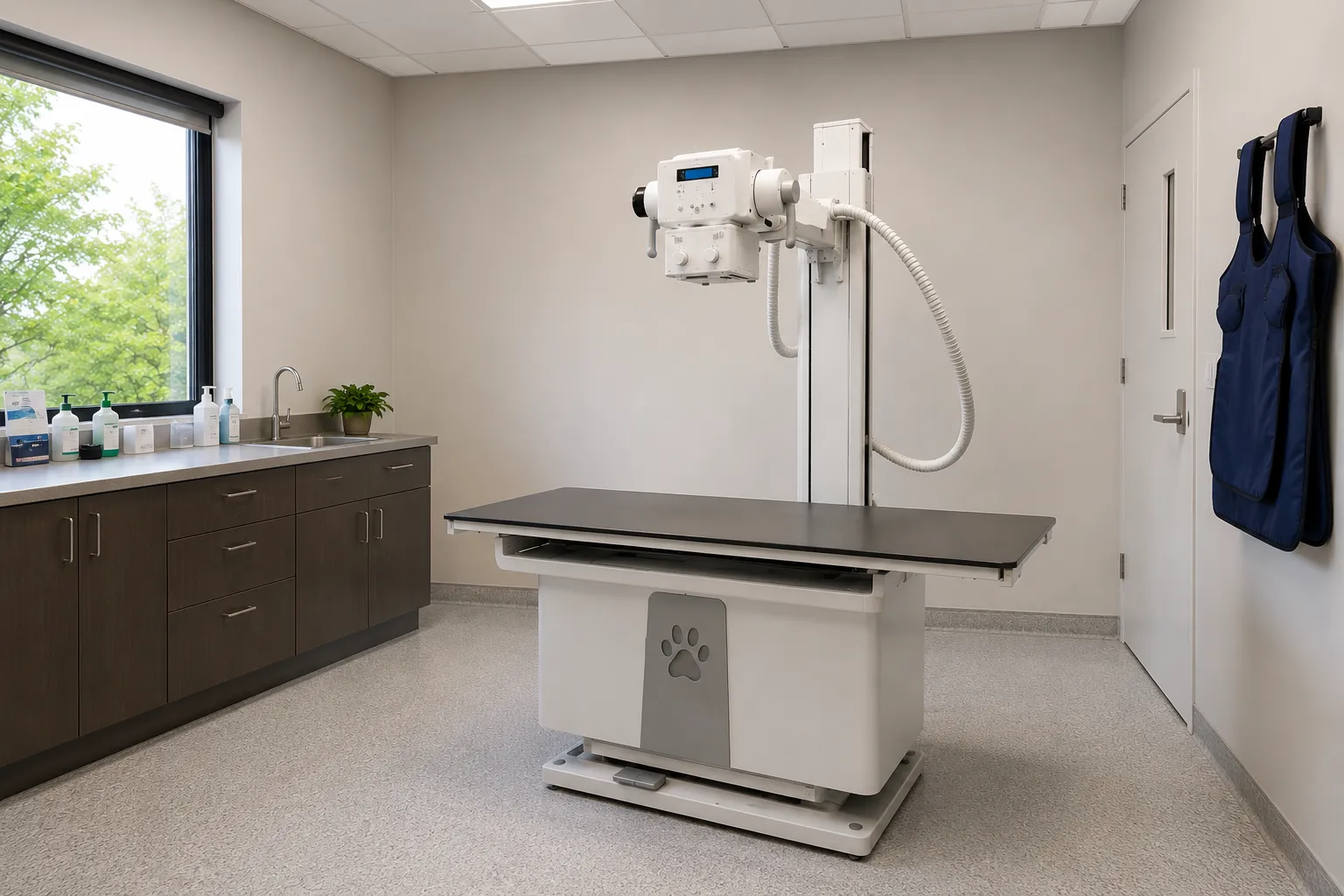
A ceiling-mounted unit with a table, the general configuration for a dedicated practice radiography room.
2. Mobile veterinary X-ray units
Wheeled units for use in different rooms or for larger patients that cannot be moved easily.
3. Portable handheld units
Compact units for field and farm work, used with strict attention to radiation safety and appropriate technique.
4. Units with a directly connected digital detector
Provide images quickly via a connector to support a workflow. Confirm the detector size for your patients.
5. Units with a computed radiography plate
Use a plate read by a reader, an established approach. Confirm the reader and plate arrangement.
6. Units with veterinary exposure settings
Include exposure settings suited to the range of animal sizes, to support images.
7. Units with positioning aids
Come with aids that help position and hold patients, improving image quality and reducing the need for people near the patient.
8. Units with dose-aware features
Support techniques that manage patients and staff. Confirm the features and how they are used.
9. Units with practice management integration
Send images to the practice's records. Confirm the connection, format and data handling.
10. Units suited to a range of patient sizes
Provide the output and detector to image small and larger patients, useful across a varied caseload.
Radiation safety, room needs and the practice
Radiation safety is central to veterinary radiography and shapes how a unit is used. A radiation protection adviser is involved in establishing arrangements, including the controlled area, local rules, protective equipment, and patient positioning, since keeping staff away from the beam matters. Positioning aids and techniques, as well as restraint during exposure, support this. The room needs the space, shielding assessed by the adviser, power, and the access the unit requires, and these should be confirmed before installation. Image handling completes the picture, since images must reach the record and any referral, so the detector, its connection and data handling should be planned. Establishing radiation safety soundly from the first examination protects the team and the patients.
What to confirm before ordering
-
Detector. Confirm the detector type and size suit your patients and workflow.
-
Positioning. Establish positioning aids that reduce manual restraint.
-
Exposure settings. Confirm settings suit the range of animal sizes.
-
Radiation safety. Verify controlled area, shielding and local rules with a radiation protection adviser.
-
Room. Confirm space, power and access for the unit.
-
Image handling. Establish record integration and data handling.
Commissioning, training and servicing
Commissioning should include radiation safety arrangements, acceptance image-quality checks, and safety-feature checks with a radiation protection adviser involved and records kept.
Training should cover positioning, exposure selection, dose-aware technique, safe holding practices and image handling, so radiography is performed safely and produces useful images.
For servicing, agree planned maintenance, image quality checks, radiation safety checks, detector maintenance and electrical safety testing. Ensure the practice receives the MHRA alerts and recalls service so any field safety notices reach the veterinary team.
Detector, positioning and radiation safety should be planned with a radiation protection adviser. Practices can work through options via buy for medical organisations.
Final thought
Choose the veterinary X-ray unit, detector positioning, and image handling for a small-animal case, with small-animal radiation safety established with expert input. Most practices are best served by a unit whose detector and settings suit their patients, positioning aids that reduce manual restraint, and sound radiation safety arrangements from the first examination.
This article provides general procurement guidance for veterinary practices. It is not clinical, veterinary or radiation protection advice, and it does not replace manufacturer instructions or expert advice on radiation safety.