Transcatheter valve replacement is a technique that fits a new heart valve using a catheter, without removing the diseased valve or opening the chest. A replacement valve, mounted inside a collapsible frame, is compressed onto a delivery catheter, guided through a blood vessel to the heart, and expanded into place to take over the job of the failing valve. Most widely known as transcatheter aortic valve implantation, or TAVI, it treats patients for whom open valve surgery carries too much risk, and its use has broadened over time.
For a buyer, this is a high-value implant programme wrapped around advanced imaging, hybrid facilities and a skilled multidisciplinary team. This guide explains how the procedure works, and the equipment and planning it demands, distinct from general vessel-opening or catheter kits.
How transcatheter valve replacement works
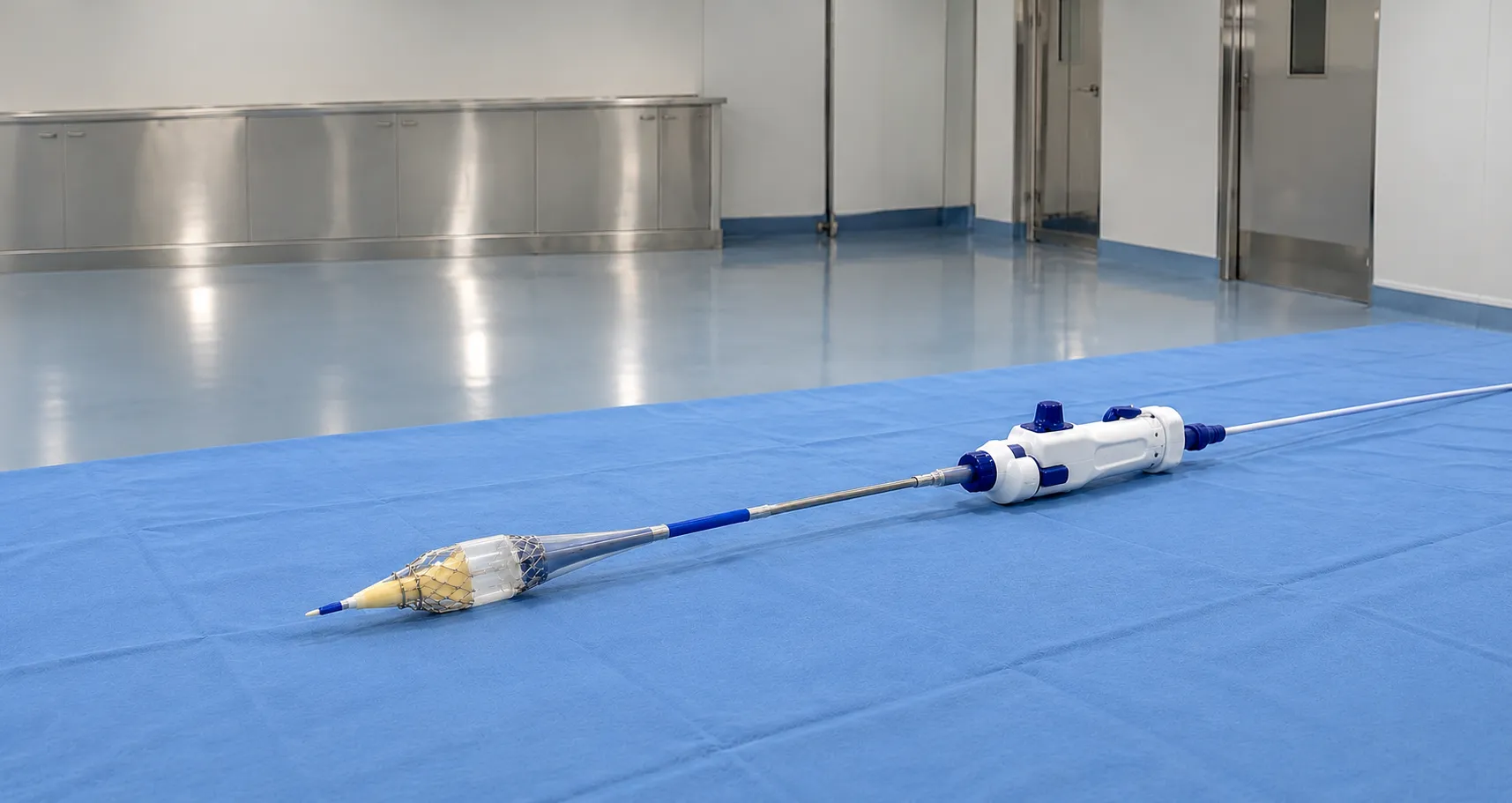
The valve prosthesis is made from biological leaflets, usually treated animal tissue, sewn into a metal frame or stent. Crimped to a small diameter, it sits on a delivery catheter advanced to the heart, most often up the aorta from the femoral artery, a route called transfemoral access. The team checks positioning with live X-ray and echocardiography, then deploys the valve precisely within the old valve, where its frame anchors it and pushes the diseased leaflets aside. The new leaflets begin working immediately, and the delivery system is withdrawn.
Accurate depth and rotational positioning matter because a valve sitting too high or too low can leak around its edge or interfere with nearby structures. That is why the deployment step is deliberate and image-guided, and why platforms that let you recapture and reposition a self-expanding valve before final release are valued in difficult anatomy. The whole sequence, from access to final deployment, is shorter than open surgery, but every stage depends on the imaging and sizing done beforehand.
Balloon-expandable and self-expanding valves
Two frame designs dominate. A balloon-expandable valve is mounted on a balloon and expanded by inflation, providing strong, controlled placement. A self-expanding valve is made from a shape-memory alloy that opens as it is unsheathed, and some designs can be recaptured and repositioned before final release. Each has different sizing, deployment behaviour, and delivery requirements, so a programme usually uses more than one platform to suit different anatomy.
Access routes and delivery systems
Transfemoral access through the groin is the default and least invasive route. When the leg arteries are too small or diseased, surgeons use alternative routes, reaching the heart through other vessels or a small chest incision. The delivery system, catheter, sheath, and deployment mechanism that carry and release the valve are specific to each valve platform, so they are purchased as a matched set. The introducer sheath size determines whether the transfemoral route is feasible for a given patient, so sizing is decided during planning.
Imaging and pre-procedure planning
Precise sizing is what makes these procedures work, and it depends on imaging. A CT angiogram of the heart and access vessels measures the valve annulus, selects the prosthesis size, and confirms the femoral arteries can accommodate the delivery sheath. During the procedure, high-quality fluoroscopy and echocardiography confirm position before and after deployment. The imaging environment is therefore a hybrid theatre or catheter laboratory with fixed high-specification X-ray, and the estate must support radiation protection to UK standards. Clinical selection follows national guidance, and the NICE and NHS set out how these services are commissioned and delivered.
Anaesthesia and recovery model
How a case is anaesthetised affects the equipment and the pathway. Many transfemoral procedures are now done under conscious sedation with local anaesthesia rather than general anaesthesia, which shortens recovery and can support a shorter hospital stay. However, more complex cases or alternative access routes may still require general anaesthesia and transoesophageal echocardiography. From a procurement standpoint, this influences the monitoring, airway and echo equipment the room must have available, and the recovery capacity the service plans for. Confirming the intended anaesthetic model early with the clinical team prevents the equipment list from being specified for the wrong pathway.
The heart team and service requirements
A multidisciplinary heart team of cardiologists, cardiac surgeons, imaging specialists, and anaesthetists runs transcatheter valve programmes, with immediate access to cardiac surgical backup if conversion to open surgery is needed. From a procurement view,, this means the implant cannot be considered in isolation: it sits within a facility, a tteammand a safety net tthat must already exist Valve platforms are premium implants supplied with their matched delivery systems, and each is a sterile, single-use, individually traceable device.
Beyond the aortic valve
Although aortic valve replacement is the established and most common transcatheter procedure, the same catheter-based principle is being applied to other heart valves, including repair and replacement approaches for the mitral and tricuspid valves. These are less standardised, involve different anatomy and delivery challenges, and use their own dedicated device systems. For a buyer, the practical point is that a structural heart programme may grow beyond aortic work over time, so it is worth understanding a supplier's wider platform range and the imaging and planning each additional procedure demands before committing to a single-purpose setup.
Consumables and the wider procedure set
The valve and its delivery system are the headline item.e m Still, each case consumes a supporting set of endovascular kit: large-bore access sheaths, guidewires stiff enough to carry the delivery system, a balloon for pre-dilatation or valve expansion, contrast, and a vessel closure device for the access site. Because the femoral access is large, closure and haemostasis matter, and the associated devices belong in the cost model. Treating the procedure as a complete consumable pathway, rather than pricing the valve alone, gives a realistic per-case figure and avoids surprises when the supporting inventory is ordered.
Standards, traceability and total cost of ownership
As implanted active-adjacent devices, transcatheter valves and their delivery systems must carry valid UKCA or CE marking under current UK rules, with the MHRA overseeing regulation. Every implant needs batch and serial recording against the patient and entry into the relevant registry, both for recall management and outcome tracking. Because unit costs are high and you must have each valve size available when a case is booked, consignment stock and agreed replenishment are common. Model whole-life cost around case volume, the size range you must hold, delivery-system consumption and imaging capacity, not the valve price alone. Your procurement team should confirm platform range, sizing support and traceability with each supplier.
Procurement checklist
-
Confirm the hybrid theatre or cath lab imaging meets the procedure's needs.
-
Decide which valve platforms, balloon-expandable or self-expanding, you support.
-
Match valve and delivery-system size ranges to your patient population.
-
Check introducer sheath sizes and access-route options are covered.
-
Verify UKCA or CE marking, MHRA registration and registry entry.
-
Put batch and serial traceability in place for each patient.
-
Agree consignment stock, sizing support and resupply with suppliers.
Conclusion
Transcatheter valve replacement is an implant programme built on precise CT sizing, hybrid imaging, matched delivery systems, and a skilled heart team, so the buying decision extends far beyond the valve itself. Plan the facility, platform range and traceability together, and price based on your real caseload and the full size inventory you must hold. To discuss sourcing valve programme consumables and equipment for a UK service through vetted suppliers, contact MediGear.
Disclaimer
This article is for informational purposes only. It is published by MediGear (medigear.uk) for general information and procurement guidance. It is not clinical, diagnostic, treatment, technical, engineering, legal or regulatory advice, nor a product endorsement, guarantee or substitute for professional assessment. MediGear does not provide medical consultations. Buyers should consult their clinical, biomedical, estates and regulatory contacts, and the manufacturer's documentation, and independently verify all specifications, certifications, compatibility and suitability before purchase. Specifications, certifications and availability are correct at the time of publication and may change without notice. MediGear is a medical-equipment distributor and does not sell medicines or pharmaceutical products.