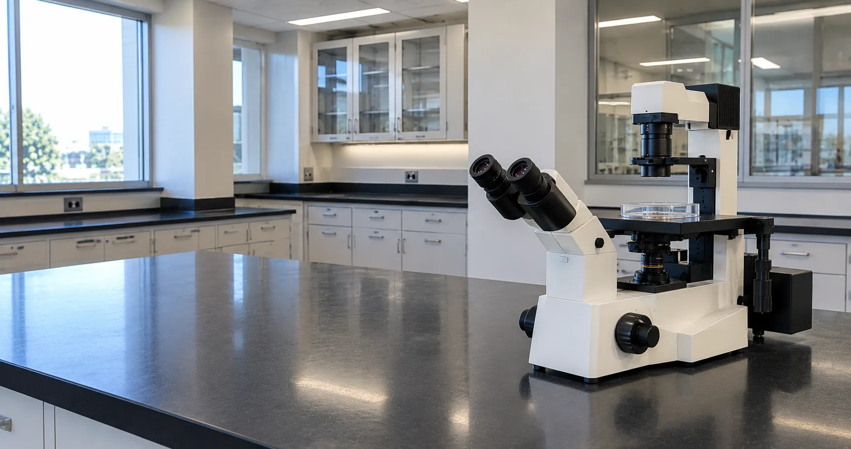
An inverted microscope is a compound microscope built upside down relative to a conventional instrument: the objective lenses sit below the stage and the light source and condenser are mounted above it, looking down through the specimen. That geometry exists for one main reason — to view living cells growing on the bottom of culture flasks, dishes and multiwell plates, through the vessel base, without disturbing the culture. For a laboratory buying its first tissue-culture microscope, the inverted layout is what makes routine checks of adherent cells, confluence and contamination possible while the cells stay in their medium.
On an upright microscope, the objectives approach the specimen from above, which forces you to place a thin sample on a slide close to the lens. That is impossible with a flask of cells sitting under a column of medium. Turning the optics under the stage solves it: the objective looks up through the vessel base at cells attached to the inside floor, with the culture and its medium safely above.
Why the Inverted Geometry Matters for Cell Culture
Cells in culture grow attached to the bottom surface of a vessel, submerged in several millimetres of medium. Only an inverted microscope can bring an objective close to that surface from underneath. The result is that cultures can be examined repeatedly, non-destructively and under sterile conditions — the vessel lid stays on, nothing touches the medium, and the flask goes straight back to the incubator. This is the workhorse of tissue culture, cell-line maintenance, assisted reproduction and any live-cell work.
Long Working-Distance Optics and Condenser
Because the objective must focus through the thickness of the vessel base and reach cells the far side, inverted microscopes use long working-distance (LWD) objectives designed to correct for the plastic or glass the light passes through. The condenser above the stage is likewise a long working-distance unit, mounted high enough to clear tall flasks and multiwell plates. Objective magnifications are usually modest — 4×, 10×, 20× and 40× cover almost all culture work — because the aim is to survey monolayers and morphology rather than resolve fine sub-cellular detail.
Contrast for Unstained Living Cells
Cultured cells are nearly transparent, so an inverted microscope is normally fitted with a contrast method that reveals them without stains. Phase contrast is the standard, turning tiny differences in optical path into visible brightness so unstained cells show clear outlines. Some instruments offer relief-contrast or modulation-contrast systems that give a shaded, three-dimensional look through plastic vessels. A companion article covers the optics of phase contrast and fluorescence in depth; here the point is that the inverted stand is the platform these methods are mounted on for live cells.
Fluorescence and Imaging Ports
Many inverted microscopes accept an epifluorescence attachment for labelled live-cell work, with an illuminator, filter cubes and often LED excitation. A trinocular head with a camera port lets you record images and video of dividing or migrating cells, and motorised versions integrate with imaging software for time-lapse. If documentation or quantitative imaging is on your roadmap, specify the camera port and software compatibility now rather than retrofitting.
Live-Cell Environmental Control
To watch cells over hours rather than minutes, the microscope must keep them alive on the stage. Stage-top incubators and enclosure chambers hold 37°C, around 5% carbon dioxide and controlled humidity so cultures behave normally during time-lapse. This turns an inverted microscope into a live-cell imaging platform for growth, migration and drug-response studies. If long-term imaging is intended, confirm the stage accepts the incubation hardware and the vessel formats you use.
Vessel Compatibility and Stage
An inverted microscope earns its place by handling the vessels your laboratory actually uses: T-25, T-75 and T-175 flasks, 35 mm and 60 mm dishes, chamber slides and 6-, 12-, 24-, 96- and 384-well plates. Universal or interchangeable stage inserts hold each format flat and stable. Check the maximum vessel height the condenser will clear and that the stage travel and inserts match your consumables, because a stage that cannot hold a plate flat wastes the optics.
Illumination, Ergonomics and Settings
Most current instruments use LED illumination, which runs cool, lasts for years and holds a stable colour temperature — helpful next to a culture hood where heat and lamp changes are unwelcome. Low, tilting binocular heads reduce neck strain during long screening sessions. Inverted microscopes are used at the bench, inside or beside a Class II safety cabinet in busy tissue-culture suites, in IVF laboratories and in research imaging rooms, so a compact footprint and simple, wipe-clean surfaces matter. You can discuss format and accessory choices with the MediGear team before ordering.
Inverted Versus Upright at a Glance
The choice between an inverted and an upright microscope follows the sample, not preference. An upright instrument, with objectives above the stage, suits thin specimens on slides — stained histology sections, blood films, fixed smears — where the coverslip sits close to the lens. An inverted instrument suits anything held in a vessel that must be viewed from below: live cultures, cells in medium, larger samples and dishes. A histology or haematology bench is typically served by an upright; a tissue-culture or IVF laboratory needs an inverted. Some departments run both, matching each to its task rather than forcing one to do everything.
Setting Up for Even Illumination
Getting clean, even contrast from an inverted microscope depends on correct Köhler illumination: setting the field and condenser apertures so the sample is lit uniformly and glare is minimised. For phase contrast the condenser phase annulus must be centred to the objective's phase ring, and each objective magnification has its own matching annulus setting. Time spent on this alignment is repaid in clearer images and more reliable assessment of cell morphology and confluence, and it is a routine part of operator training in a busy culture suite.
Standards and Decontamination
As laboratory instruments, inverted microscopes should carry UKCA or CE marking with electrical safety to the IEC 61010-1 family. Because they live in tissue culture settings, near category-2 organisms, surfaces must tolerate routine disinfection; check that the manufacturer approves your wipe or spray, and that objectives and stage inserts can be cleaned without damaging coatings. Guidance on device regulation is published by the MHRA.
Procurement Checklist
Before choosing an inverted microscope, verify the following:
-
Objective set (typically 4× to 40×) as long-working-distance types corrected for your vessel bases.
-
Contrast method — phase contrast or relief contrast — suited to unstained cells in plastic vessels.
-
Vessel compatibility, with stage inserts for the flasks, dishes and plate formats you run.
-
Condenser working distance tall enough to clear your largest flasks and plates.
-
Fluorescence and camera ports if labelled imaging or documentation is planned.
-
Stage-top incubation compatibility for live-cell time-lapse, if required.
-
Cleanable surfaces and disinfectant compatibility for tissue-culture use.
-
UKCA or CE marking, warranty, servicing and objective availability.
Total Cost of Ownership
Inverted microscopes are relatively economical to run. LED illumination removes lamp replacement, and a basic phase-contrast culture stand needs little beyond occasional objective cleaning. Costs rise with fluorescence, motorisation, cameras and stage incubation, and those accessories often outweigh the base microscope. Buy the stand with the ports and stage you will need over its life, standardise objective magnifications across the department for shared training, and you will keep both capital and running costs predictable. Registering standing requirements through our buyer services helps match a specification to your workflow.
Conclusion
An inverted microscope puts the objectives beneath the stage so it can look up through a flask or plate at living cells attached to the vessel floor, which is why it is the standard tool of tissue culture, IVF and live-cell imaging. Match objectives, contrast, vessel formats and any fluorescence or incubation to your workflow, and confirm cleanability and marking. To compare inverted microscopes and accessories for your culture suite, contact MediGear.
Disclaimer
This article is for informational purposes only. It is published by MediGear (medigear.uk) for general information and procurement guidance, and is not clinical, diagnostic, treatment, technical, engineering, legal or regulatory advice, nor a product endorsement, guarantee or substitute for professional assessment. MediGear does not provide medical consultations. Buyers should consult their clinical, biomedical, estates and regulatory contacts, and the manufacturer's documentation, and independently verify all specifications, certifications, compatibility and suitability before purchase. Specifications, certifications and availability are correct at the time of publication and may change without notice. MediGear is a medical-equipment distributor and does not sell medicines or pharmaceutical products.