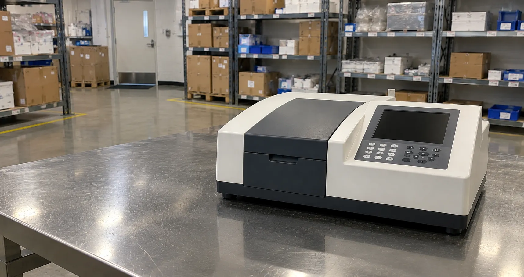
A spectrophotometer measures how much light a sample absorbs at each wavelength by passing a beam through the sample and comparing the transmitted intensity with a reference. Absorbance rises with the concentration of the absorbing substance, so the instrument turns a colour or ultraviolet signature into a number, and that number underpins quantitative work across clinical chemistry, molecular biology and water testing.
The physics is the Beer-Lambert law, and the buying decision turns on wavelength range, optical design and the sample format you use. This overview explains how absorbance is measured, the main instrument types, and the specifications a laboratory should compare before ordering.
The Beer-Lambert Law
Absorbance (A) equals the molar absorptivity of the substance multiplied by the path length and the concentration: A = εcl. Because path length and absorptivity are fixed for a given measurement, absorbance is directly proportional to concentration, which is why a spectrophotometer can quantify an analyte from a single reading once a calibration or known absorptivity is established. The relationship holds only over a limited absorbance range; above roughly 1.5 to 2 A, stray light and detector limits cause it to curve, so samples are diluted to stay in the linear region.
Inside the Instrument
A UV-Vis spectrophotometer has five functional parts. A light source provides the beam, typically a deuterium lamp for the ultraviolet (below about 350 nm) and a tungsten-halogen lamp for the visible and near-infrared. A monochromator, usually a diffraction grating, selects a narrow band of wavelengths. The beam passes through the sample in a cuvette, and a detector, a photodiode, photomultiplier or array sensor, measures the transmitted light. Electronics convert transmittance to absorbance and display the result.
Instrument Designs Compared
Single-beam
The simplest design measures a blank, then the sample, through one light path. It is economical and sensitive but relies on a stable source and periodic blanking.
Double-beam
The beam is split between sample and reference so lamp drift is corrected in real time, which suits long kinetic runs and demanding quantitative work.
Diode-array
Here the sample is illuminated with full-spectrum light and a fixed grating disperses the transmitted beam onto an array detector, capturing the whole spectrum in a fraction of a second. Diode-array instruments are fast and robust, ideal for scanning and for detectors on liquid-chromatography systems.
Wavelength Range and Optical Quality
Range decides what you can measure: a visible-only instrument (typically 320 to 1100 nm) handles colourimetric assays, while UV-Vis (around 190 to 1100 nm) is needed for nucleic acids and proteins that absorb in the ultraviolet. Three optical specifications separate models. Spectral bandwidth (slit width) governs how well closely spaced peaks are resolved; wavelength accuracy affects whether you read a peak at its true maximum; and stray light sets the highest absorbance you can trust. For pharmacopoeial and nucleic-acid work,k these are not optional extras but acceptance criteria.
Cuvettes and Sample Formats
The standard sample holder is a cuvette with a 10 mm path length. Material matters: optical glass and disposable plastic cuvettes work in the visible range, but only quartz transmits ultraviolet, so DNA and protein measurements below 300 nm require quartz. Micro-cuvettes reduce the volume needed, and low-volume adaptors let femtoliters be measured.
Micro-volume and nanovolume
Dedicated micro-volume instruments hold a 1 to 2 µL drop between two pedestals with a very short path length, reading concentrated DNA, RNA or protein samples without dilution or cuvettes. The A260/A280 ratio from such a reading indicates nucleic-acid purity, which is why these units are a molecular-biology staple. Some models combine a pedestal and a cuvette port, so one instrument covers both formats.
Common Applications
Spectrophotometers quantify nucleic acids and proteins, follow enzyme kinetics by reading absorbance over time, run colourimetric and ELISA-style endpoint assays, and support water and environmental testing. In clinical chemistry,y the same principle drives analyser photometry. Because each application has its own wavelength and sensitivity demands, the assay list should define the instrument rather than the reverse; our buyer services can help match a model to a workload.
Reading Modes and Software
The measurement modes an instrument offers often decide whether it fits a workflow. Fixed-wavelength reading gives a single absorbance for endpoint assays; multi-wavelength reading takes several set points at once, as in the A260/A280 nucleic-acid check; spectrum scanning records absorbance across a range to find or confirm a peak; and kinetic mode logs absorbance against time to derive an enzyme rate. Quantitation mode builds a standard curve from known concentrations and reads unknowns against it. A dedicated nucleic-acid or protein application may pre-set the wavelengths and calculations, saving method-building, so check that the software covers the assays you run rather than assuming it does. Colourimeters and Filter Photometers
Not every measurement needs a full spectrophotometer. A colourimeter or filter photometer uses fixed optical filters instead of a scanning monochromator to read one or a few wavelengths, and for a routine single-wavelength colourimetric test it is cheaper, rugged and simple. The trade-off is flexibility: a filter instrument cannot scan a spectrum or read arbitrary wavelengths, so it suits a fixed assay rather than a general laboratory. Deciding between the two comes down to whether your assays are fixed and few, or varied and evolving.
Handling also affects the numbers. Cuvettes must be filled above the beam height, wiped clear of fingerprints, and oriented the same way each time, since scratches and smears add spurious absorbance. Instruments need a short warm-up for the source to stabilise, and a fresh blank of the same matrix as the sample keeps the baseline honest.
Calibration, Standards and Regulation
Performance is verified against reference standards. Wavelength accuracy is checked with holmium oxide or didymium filters that have known absorption peaks, photometric accuracy with neutral-density or potassium dichromate standards, and stray light with cut-off solutions, ideally with traceable certification. Laboratories working to pharmacopoeial methods follow defined qualification procedures, and where a spectrophotometer forms part of a diagnostic workflow, its regulatory status can be confirmed through the MHRA. Keep a calibration log and run the checks at the interval your quality system sets.
Procurement Checklist
Before buying a spectrophotometer, confirm the following:
-
Wavelength range (visible-only versus full UV-Vis) matched to whether you measure nucleic acids and proteins or only coloured solutions.
-
Optical design (single-beam, double-beam or diode-array) appropriate to kinetics, scanning or routine endpoint work.
-
Spectral bandwidth, wavelength accuracy and stray light, checked against your method or pharmacopoeial acceptance criteria.
-
Sample format: standard cuvette, micro-cuvette or micro-volume pedestal, and whether quartz cuvettes are needed for UV.
-
Calibration standards and qualification support, with traceable holmium and photometric references.
-
Software and connectivity for data capture, audit trails and LIMS or chromatography integration.
-
Lamp life and replacement cost for the deuterium and tungsten sources, plus service availability.
Total Cost of Ownership
The lamps are the main consumable: a deuterium source has a finite rated life and dims with use, while tungsten-halogen lamps are cheaper but also age. Quartz cuvettes are durable but costly and easily scratched, whereas disposable plastic cuvettes suit routine visible work. Budget for calibration standards, periodic qualification and a service contract, and standardise cuvette formats across the laboratory to simplify stocking.
Conclusion
A spectrophotometer applies the Beer-Lambert law to turn light absorption into concentration, and specifying one comes down to wavelength range, optical design and sample format. Decide whether you need ultraviolet reach, diode-array speed or a micro-volume pedestal, then hold the instrument to defined calibration standards. To compare UV-Vis and micro-volume models or arrange qualification support, contact the MediGear team and set out your assay requirements.
Disclaimer
This article is for informational purposes only. It is published by MediGear (medigear.uk) for general information and procurement guidance, and is not clinical, diagnostic, treatment, technical, engineering, legal or regulatory advice, nor a product endorsement, guarantee or substitute for professional assessment. MediGear does not provide medical consultations. Buyers should consult their clinical, biomedical, estates and regulatory contacts, and the manufacturer's documentation, and independently verify all specifications, certifications, compatibility and suitability before purchase. Specifications, certifications and availability are correct at the time of publication and may change without notice. MediGear is a medical-equipment distributor and does not sell medicines or pharmaceutical products.