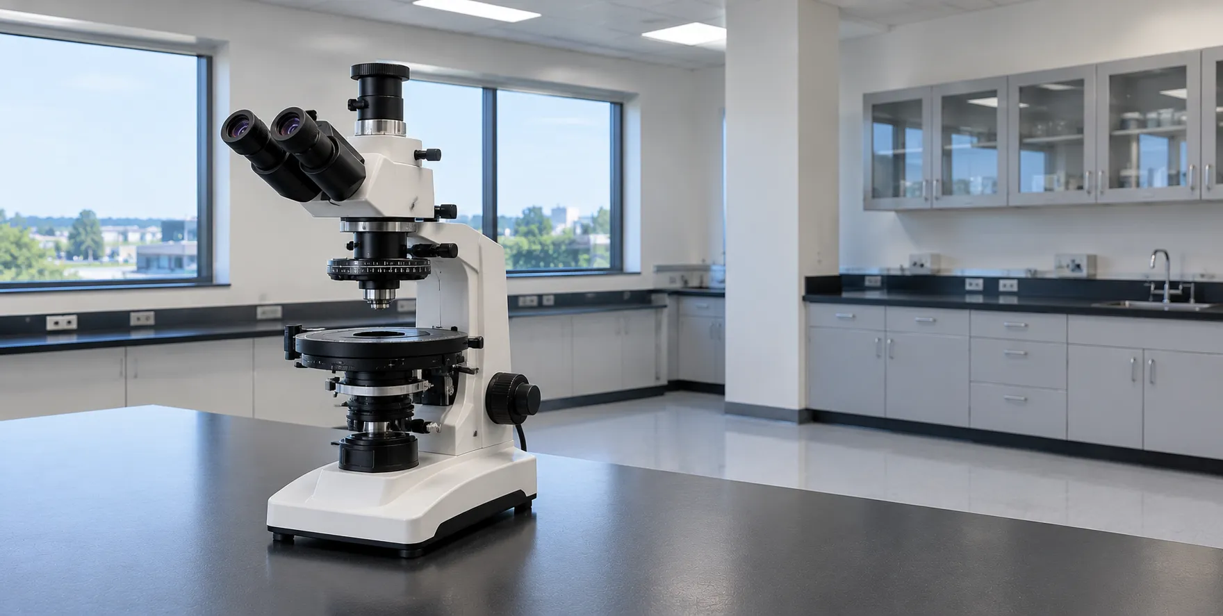
A polarising microscope identifies materials by how they interact with polarised light, using two polarising filters — a polariser below the stage and an analyser above it — set at right angles so the field is dark until a birefringent object rotates the light and lights up against black. That single property, birefringence, is what makes the instrument indispensable for identifying crystals: in clinical pathology it distinguishes gout from pseudogout in joint fluid, and in mineralogy and materials work it identifies crystalline phases, fibres and strain. For a laboratory buyer, the value lies in the strain-free optics and rotating stage that reveal these effects reliably.
The principle is straightforward. Light from the polariser vibrates in one plane. A non-birefringent (isotropic) material leaves it unchanged, so the crossed analyser blocks it and the object stays dark. A birefringent (anisotropic) crystal splits and retards the light into two components; when they recombine at the analyser, some light passes, and the crystal appears bright, often in interference colours, on a black background.
Crossed Polarizers and Birefringence: The polarizers at the microscope are the crossed polarizer and analyser. With no birefringent object between them, the field is extinguished. A birefringent crystal has two refractive indices; the difference between them, multiplied by the crystal thickness, sets the optical retardation and therefore the interference colour seen between crossed polarisers. Rotating the specimen changes brightness through positions of extinction and maximum brightness every 90°, which itself is diagnostic. This is a qualitatively different mechanism from dark-field or phase contrast, and it reports specifically on molecular and crystalline order.
The Compensator and Sign of Elongation
A key accessory is the first-order red (lambda) compensator, a retardation plate inserted at 45° in the light path. It adds a known, fixed retardation, shifting the background to magenta and colouring birefringent objects. Whether a crystal appears blue or yellow when its long axis is aligned with the compensator's slow axis tells you the sign of birefringence — positive or negative — which is the deciding test in crystal identification. This is exactly how clinical laboratories separate the two common joint-fluid crystals.
Clinical Crystal Identification
In synovial (joint) fluid analysis, compensated polarised light distinguishes the crystals of two conditions. Monosodium urate crystals, seen in gout, are needle-shaped and strongly negatively birefringent: yellow when their long axis lies parallel to the compensator's slow axis, blue when perpendicular. Calcium pyrophosphate crystals, seen in pseudogout (calcium pyrophosphate deposition), are rhomboid or rod-shaped and weakly positively birefringent, showing the opposite colours. The colour-versus-orientation rule, read with the red compensator, is what separates them. The instrument also demonstrates the apple-green birefringence of Congo-red-stained amyloid and helps identify fibres such as asbestos.
Strain-Free Optics and the Rotating Stage
Polarising work demands components that do not themselves alter polarised light. Objectives and condensers are made strain-free, because internal stress in ordinary glass would introduce its own birefringence and false colour. A graduated rotating stage, marked in 360° with a vernier, lets the specimen be turned to find extinction and measure angles. A centring mechanism keeps the object on the axis of rotation. These features distinguish a true polarising microscope from a standard one with a pair of filters clipped on.
Conoscopy and the Bertrand Lens
Beyond ordinary (orthoscopic) viewing, a polarising microscope can work in conoscopic mode. A Bertrand lens brings the interference figure at the back focal plane of the objective into view, revealing whether a crystal is uniaxial or biaxial and its optic sign. Mineralogists and materials scientists use this to characterise unknown crystals; clinical laboratories rarely need it, so decide whether conoscopy is worth specifying for your work.
Illumination and Documentation
A stable, even light source is essential because interference colours depend on it; modern instruments use LED illumination with consistent colour temperature. A trinocular head and camera port let laboratories photograph crystals for records and reporting, which matters where results feed into patient notes or quality assurance. Digital documentation also supports training and second opinions, and captured images help resolve difficult cases without recalling the original preparation. For crystal work the light must be intense enough to reveal weakly birefringent objects yet colour-stable so interference colours are read consistently, which is why an even, well-regulated source is not a minor specification. Standing requirements can be registered through our buyer services.
Measuring Extinction and Interference Colours
Two measurements make the polarising microscope quantitative rather than merely descriptive. The extinction angle — the stage rotation between a crystal's reference edge and the position where it goes dark — is read on the graduated stage and is characteristic for many crystals. The interference colour a birefringent object shows between crossed polarisers, compared against a standard Michel-Levy chart, relates the retardation to the crystal's thickness and birefringence, helping identify the material. A vernier on the rotating stage and a well-calibrated compensator are what make these readings repeatable between operators, which is why the mechanical quality of the stage is not a trivial detail.
Beyond the Joint-Fluid Clinic
Crystal identification reaches well past synovial fluid. In tissue pathology the microscope confirms the apple-green birefringence of Congo-red-stained amyloid, and it identifies foreign material and crystalline deposits in sections. In occupational and environmental work it helps identify asbestos and other mineral fibres by their optical signs. Pharmaceutical and chemical laboratories use it to distinguish polymorphs and to check crystallinity, and geology and materials science rely on it to identify minerals in thin section. A single well-specified instrument therefore serves clinical, industrial and research users, provided its optics and accessories suit the most demanding of those tasks.
Standards, Regulation and Care
As a laboratory instrument, the microscope should carry UKCA or ECE marking with electrical safety to the IEC 61010-1 family, and where results are used, the laboratory works within its own accreditation framework. Polarising optics need careful handling: the polariser and analyser films degrade with heat and ultraviolet exposure, so avoid overpowered lamps and store the instrument out of strong light. Clinical guidance on conditions such as gout is published by the National Institute for Health and Care Excellence, and device-regulation guidance by the MHRA.
Procurement Checklist
Before choosing a polarising microscope, confirm the following:
-
Strain-free objectives and condenser genuinely intended for polarised light, not standard optics.
-
Rotatable graduated stage with centring and a vernier for measuring extinction angles.
-
First-order red (lambda) compensator and slots for additional retardation plates.
-
Analyser and polariser quality, with the analyser rotatable or fixed at a true 90°.
-
Bertrand lens and conoscopy if you need to characterise unknown crystals.
-
Magnification range suited to your work, from low-power survey to oil-immersion detail.
-
Camera port and documentation for reporting and quality assurance.
-
UKCA or CE marking, warranty, servicing and spare optics.
Total Cost of Ownership
A polarising microscope is a modest running cost once bought: LED illumination removes lamp replacement, and the main consumable is care of the polarising films and occasional objective cleaning. The larger decisions are one-off — strain-free optics, the compensator set and conoscopy hardware — so specify these correctly at purchase rather than upgrading piecemeal. Standardising the compensator convention and reporting workflow across the laboratory keeps results consistent between operators.
Conclusion
A polarising microscope reads birefringence through crossed polarisers, a rotating stage and a first-order red compensator, which is why it identifies crystals from urate in gout to minerals and fibres. Specify strain-free optics, the right compensator and any conoscopy your work needs, and confirm marking and documentation. To compare polarising microscopes and accessories for crystal identification, talk to the MediGear team.
Disclaimer
This article is for informational purposes only. It is published by MediGear (medigear.uk) for general information and procurement guidance, and is not clinical, diagnostic, treatment, technical, engineering, legal or regulatory advice, nor a product endorsement, guarantee or substitute for professional assessment. MediGear does not provide medical consultations. Buyers should consult their clinical, biomedical, estates and regulatory contacts, and the manufacturer's documentation, and independently verify all specifications, certifications, compatibility and suitability before purchase. Specifications, certifications and availability are correct at the time of publication and may change without notice. MediGear is a medical-equipment distributor and does not sell medicines or pharmaceutical products.