Confocal microscopy is a fluorescence imaging method that uses a pinhole to reject out-of-focus light, capturing a thin, sharp optical slice from within a thick specimen. By blocking the blurred light coming from above and below the focal plane, it builds clean images plane by plane. It stacks them into a three-dimensional reconstruction, something conventional widefield fluorescence cannot do. That optical sectioning is the whole point of confocal, and it is why laboratories reach for it when a widefield image is too blurred to interpret.
Because confocal is itself fluorescence-based, this article does not re-explain fluorophores; a sibling piece covers widefield fluorescence. Here the focus is the pinhole, scanning, the Z-stack, and the specifications that determine which confocal to buy.
The Pinhole and Optical Sectioning
The defining component is a pinhole aperture placed at a point optically conjugate to the focal plane, hence confocal. Light emitted from the exact plane of focus is imaged onto the pinhole and passes through to the detector; light from any other depth is focused elsewhere and is largely blocked by the pinhole edge. As a result, only in-focus light is recorded, so a single confocal image is a genuine slice through the specimen rather than a projection of its whole thickness. The pinhole diameter, set in Airy units, trades resolution and optical-section thickness against signal: a smaller pinhole gives thinner sections but dimmer images.
Laser Scanning Confocal
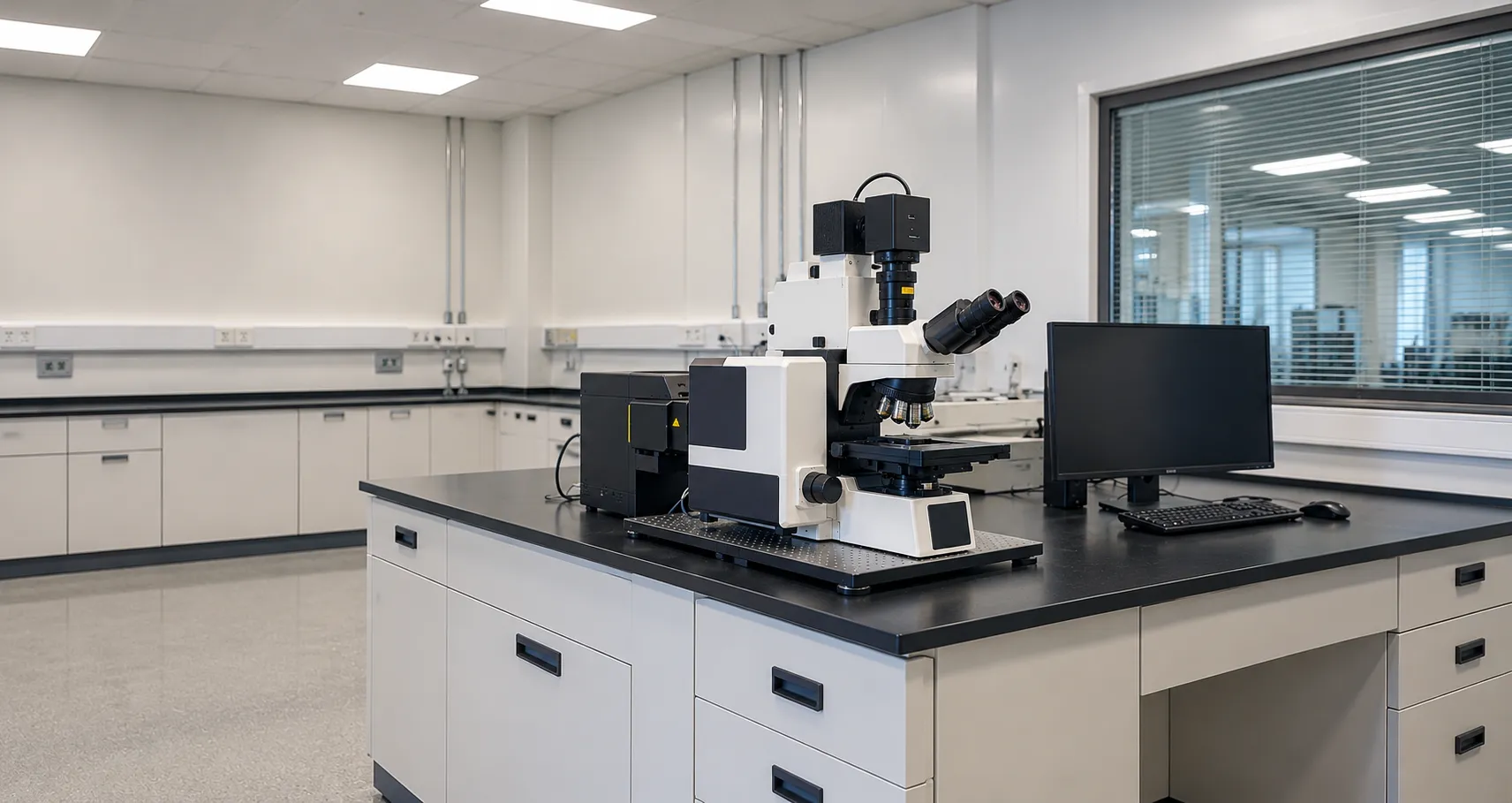
The most common form is the laser scanning confocal microscope. Because the pinhole only passes light from one point, the instrument must build the image point by point: galvanometer mirrors sweep a focused laser spot across the field, and the emission from each point is measured in turn, then assembled into a full frame. Lasers provide the intense, single-wavelength excitation this demands, commonly at 405, 488, 561 and 640 nm to match widely used fluorophores. Detection uses sensitive photomultiplier tubes or GaAsP detectors rather than a camera, because the signal arrives one point at a time.
Building a Z-Stack and 3D Image
Because each image is a thin slice, moving the focus in small, precise steps captures a series of slices at different depths, a Z-stack. Software then reconstructs the stack into a three-dimensional view, allowing researchers to measure structures in depth, rotate them, and render them. Adding time gives four-dimensional imaging of living samples, and scanning the emission wavelength gives spectral data to separate overlapping dyes. This ability to optically section and rebuild in three dimensions, without physically cutting the specimen, sets confocal apart from widefield imaging.
Spinning Disk Confocal
Point-by-point scanning is thorough but slow, which is a problem for fast living processes and can bleach or damage delicate cells. Spinning disk confocal solves this with a rotating disk carrying many pinholes (often a Nipkow disk paired with a microlens array), so it scans thousands of points in parallel, and a camera captures whole frames at high speed. The spinning disk trades some optical-sectioning strictness for much faster, gentler imaging, making it the usual choice for live-cell work, while laser scanning suits fixed samples and the thickest specimens.
Resolution and Section Thickness
Confocal improves both lateral and axial resolution over widefield, and the closed pinhole gives a modest lateral gain in addition to its main axial benefit. Section thickness depends on the objective numerical aperture, wavelength, and pinhole setting, so a high-NA oil objective with the pinhole near one Airy unit gives the thinnest, sharpest slices. Buyers should think of the objective and pinhole together, because a low-NA objective cannot deliver thin sections, no matter how the pinhole is set.
What Confocal Adds Over Widefield
Widefield fluorescence collects light from the entire depth of the sample at once, so thick specimens look foggy with out-of-focus haze. Confocal removes that haze by rejecting out-of-focus light at the pinhole, which is critical for thick tissue, embryos, biofilms, and any three-dimensional sample. The trade-off is complexity, cost, and slower acquisition, so you choose confocal when optical sectioning is genuinely needed rather than as a default; for thin monolayers, a widefield system is often enough. Our buyer services can help weigh the two against a real workload.
Scan Speed, Dwell Time and Averaging
On a laser scanning system, the image is built one pixel at a time, so several settings trade image quality against speed. Pixel dwell time, how long the laser rests on each point, sets how much signal is collected: longer dwell gives a brighter, cleaner image but a slower frame and more bleaching. Frame averaging combines several scans to suppress noise at the cost of time and exposure. Bidirectional scanning, sampling on both sweeps of the mirror, roughly doubles speed. Choosing sampling fine enough to capture the resolution the optics can deliver, without over-sampling and wasting light, is part of getting the best from the instrument, and it explains why a confocal is slower than a camera-based widefield capture.
Applications and Related Techniques
Confocal earns its place wherever depth matters: imaging thick tissue sections, tracking labelled structures in three dimensions, measuring whether two molecules occupy the same location (colocalisation), and following dynamics in living cells over time. Several methods extend the same idea. Multiphoton microscopy uses long-wavelength pulsed lasers to reach deeper into tissue with less damage, while super-resolution add-ons push detail below the classical diffraction limit. These are specialist extensions; for most laboratories, the decision is whether standard confocal sectioning answers the question, and if so, which platform delivers it.
Practical Requirements
A confocal is a system, not just a microscope. It needs vibration isolation, a stable environment, and often a live-cell chamber that controls temperature, humidity, and carbon dioxide for time-lapse work. The lasers, scanner,,r and detectors require a service contract, and the data volumes from Z-stacks and time series require substantial storage and analysis software. Scope these running requirements, alongside the purchase price, before committing.
Standards, Regulation and Servicing
Laser safety is a real consideration: confocal systems contain Class 3B or 4 lasers, and installation must meet laser-safety requirements, with the HSE informing workplace duties. If a confocal supports diagnostic reporting, the MHRA can confirm its regulatory status. A service contract covering laser alignment, detector performance and scanner calibration protects both image quality and uptime, so confirm response times and spares availability before purchase.
Procurement Checklist
Before buying a confocal microscope, confirm the following:
-
Choose laser scanning versus spinning disk based on whether you need the thinnest sections or fast, gentle live-cell imaging.
-
Laser lines (for example, 405, 488, 561 and 640 nm) matched to your fluorophores.
-
Detectors: photomultiplier or high-sensitivity GaAsP for scanning, or a fast camera for spinning disk.
-
Objectives with high numerical aperture and the correct immersion for thin optical sections.
-
Z-drive precision and the software for Z-stacks, 3D reconstruction and any time-lapse or spectral modes.
-
Environmental control such as a live-cell incubation chamber, plus vibration isolation.
-
Laser-safety compliance, service contract, and data storage included in the total cost.
Conclusion
Confocal microscopy cuts thin optical slices using a pinhole to reject out-of-focus light, then stacks those slices into three-dimensional images, its decisive advantage over widefield fluorescence. Choose laser scanning or spinning disk according to your samples, match lasers and objectives to your fluorophores and sectioning needs, and budget for laser safety, environmental control and servicing. To compare confocal platforms or scope an installation, contact the MediGear team.
Disclaimer
This article is for informational purposes only. It is published by MediGear (medigear.uk) for general information and procurement guidance. It is not clinical, diagnostic, treatment, technical, engineering, legal or regulatory advice, nor a product endorsement, guarantee or substitute for professional assessment. MediGear does not provide medical consultations. Buyers should consult their clinical, biomedical, estates and regulatory contacts, and the manufacturer's documentation, and independently verify all specifications, certifications, compatibility and suitability before purchase. Specifications, certifications and availability are correct at the time of publication and may change without notice. MediGear is a medical-equipment distributor and does not sell medicines or pharmaceutical products.