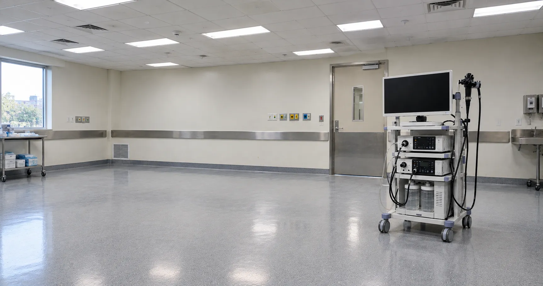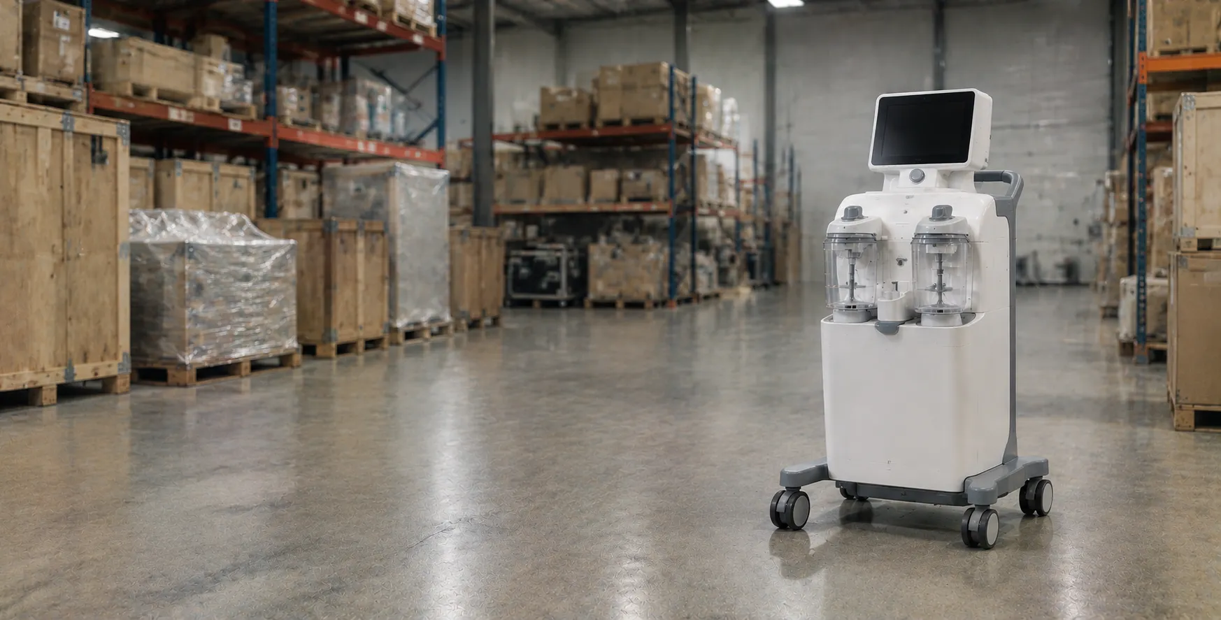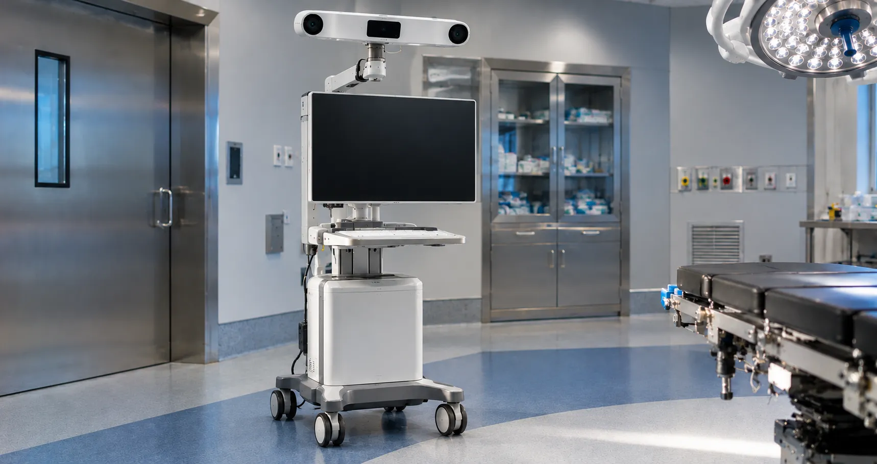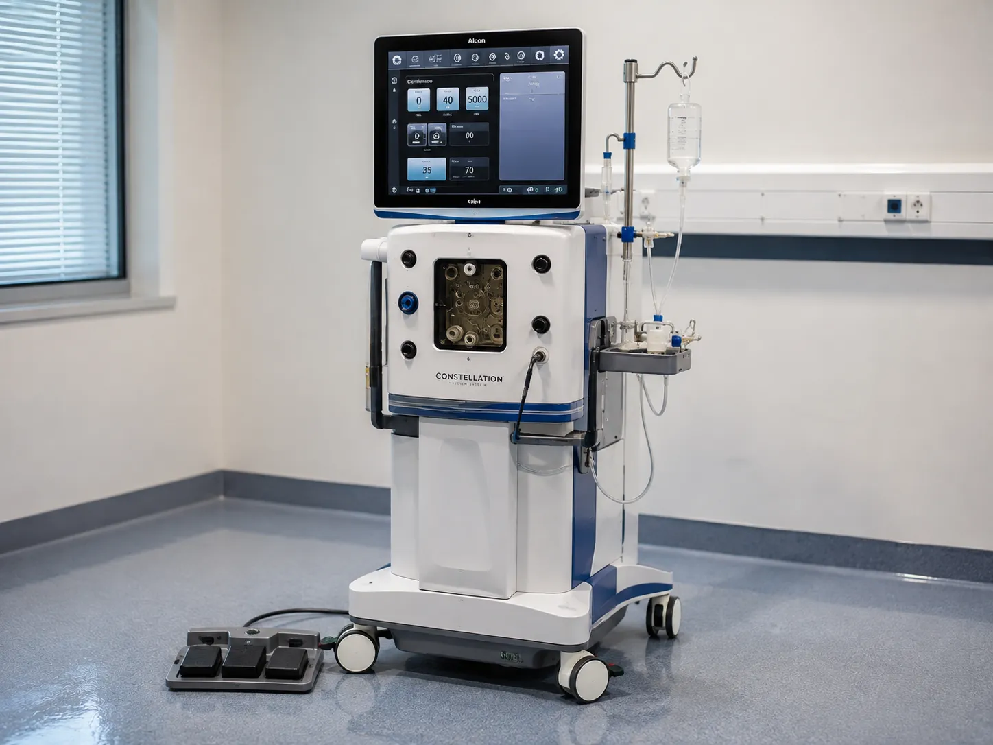
A vitrectomy machine is the surgical platform used to remove the vitreous gel from the back of the eye and to perform retinal surgery. A vitreous cutter is a fine probe with a guillotine-style port that opens and closes rapidly to nibble the gel into small pieces while aspirating them away, allowing the vitreous to be removed without tugging on the delicate retina. Around that cutter, the console coordinates aspiration. This pressurised infusion maintains the eye's shape and intraocular illumination, providing the surgeon with a controlled environment to repair conditions such as retinal detachment, macular problems and vitreous haemorrhage.
For a buyer, vitrectomy sits at the more complex end of ophthalmic capital equipment. 'It's clear how it differs from cataract surgery: phacoemulsification works at the front of the eye on the lens, whereas vitrectomy works at the back of the eye on the vitreous and retina and requires its own cutter, fluidics, and lighting.
How the vitreous cutter and fluidics work
The cutter is introduced through a small port in the sclera and its inner needle oscillates to open and close a side aperture many thousands of times a minute. Each cut takes a minute bite of vitreous, which is drawn away through the probe by the aspiration system. A high cut rate matters because faster, smaller bites reduce the traction transmitted to the retina during removal, which is why cut-rate performance is a headline specification. Older probes were driven closed and sprung open, cutting in one direction, whereas dual-action pneumatic cutters drive the port both open and closed, keeping it open longer for a given rate and improving flow at high speeds. Some probes place the port very close to the tip, allowing the surgeon to bring the vitreous closer to the retina, which matters in detachment work. At the same time, an infusion line delivers balanced fluid or, later in the case, air or gas to maintain stable pressure as the vitreous is removed. Managing infusion pressure against aspiration is the core of vitreoretinal fluidics, and modern consoles actively regulate intraocular pressure to keep the eye stable throughout.
Instrument gauges and why they matter
Vitrectomy instruments are sized by gauge, with smaller-bore systems — commonly 23, 25 and 27 gauge — allowing entry ports that are often self-sealing and need no suture, which supports faster recovery. A finer gauge means a smaller wound but a narrower lumen, so console fluidics and cut rate must be tuned to keep as maintainable as possible through aspiration withthee thinner probe as maintainable thrthegh aspiration.s a platform supports and that the full instrument set — cutters, forceps, scissors, laser probes and light pipes — is available in the gauges the surgeons prefer, because mixing gauges mid-case is not practical.
The parts of a vitrectomy platform
Console and fluidics
The capital unit houses the cutter drive, the aspiration pump, active infusion-pressure control and the interface, and it stores surgeon presets for different steps of a case.
Illumination and viewing
Endoillumination probes light the inside of the eye, and the console typically drives a bright, filtered light source; the surgeon views through the operating microscope with a wide-angle viewing system. Chandelier fixtures can be fixed into the sclera to light the entire cavity, freeing the surgeon for bimanual work—sources: xenon or LED with filters that cut the shorter, more phototoxic wavelengths. Light-source type and safety filtering are part of the specification.
Combined and adjunct functions
Many platforms combine posterior vitrectomy with anterior phacoemulsification in one con, nd integrate an endolaser and controls to deliver silicone oil or gas tamponade. Tamponade agents hold the retina in position after repair, and a viscous-fluid injection module lets the surgeon deliver and later remove silicone oil through the same platform. This breadth is attractive where a unit does mixed casework.
Key specifications a buyer must check
Confirm the maximum cut rate and the supported gauges, and check how the console controls intraocular pressure, since active pressure regulation is a significant stability feature. Establish which functions are integrated — endolaser, phacoemulsification, viscous-fluid injection for oil and gas — and whether the light source meets current safety-filtering expectations. Review the range and gauge availability of the wider instrument set, the surgeon-preset capacity and the interface. Check which components are single-use, notably the cutter probes and tubing packs, and confirm the packs are readily supplied and specific to the console. As with any theatre platform, verify the accessory connectors and foot-pedal configuration suit the team.
Standards, regulation and light safety
A vitrectomy system is an active medical device. It must carry a CA or CE marking and be designed to the general medical electrical safety standard IEC 60601-1, with the associated electromagnetic compatibility requirements. Because intraocular illumination is intense, the light source and its filtering warrant particular attention to photic-safety guidance in the manufacturer's documentation. Ask for the declaration of conformity and confirm the manufacturer's registration for the Great Britain market with the MHRA, which also receives reports of any device-related incident. Wider service and safety guidance relevant to eye services is published by the NHS and by the unit on its vitreoretinal provision. However, that guidance surrounds the device and focuses on more than forming part of it.
Decontamination and single-use considerations
Vitreoretinal cutter probes and tubing sets are single-use and discarded after each patient, which eliminates processing risk for the most intricate parts of the system. Reusable instruments such as certain forceps, scissors and light pipes, where offered, must be cleaned and sterilised strictly in accordance with the manufacturer's validated method, with careful attention to their fine tips and lumens. As with anterior-segment surgery, teams should remain alert to the risk of toxic anterior segment syndrome and other contaminant-related reactions, so follow sterile-service decontamination instructions precisely. These details should be verified against the manufacturer's documentation before a platform is adopted.
Care settings and total cost of ownership
Vitrectomy is performed in specialist ophthalmic theatres and vitreoretinal units within the NHS and the independent sector, rather than in general clinics, because it requires the console, microscope, viewing systems and a trained surgical team. The economics follow the same pattern as other energy-and-fluidics platforms: the console is a one-off capital cost, but each case consumes a single-use cutter and pack, so annual spend tracks case volume multiplied by pack price. Add service contracts, software updates, endolaser fibre and probe costs, the light-source maintenance, and the tamponade agents used per case. Where a combined console covers both phaco and vitrectomy, standardisation reduces the need for theatre training. Our team compares our platform against a unit's casework and consumable budget — register as a buyer or contact us through the contact page.
Procurement checklist
-
Confirm the maximum cut rate and the supported instrument gauges.
-
Check how the console controls intraocular pressure during a case.
-
Establish which functions are integrated, including endolaser and phaco.
-
Verify the light source meets current photic-safety filtering guidance.
-
Review the instrument set and gauge availability for your surgeons.
-
Confirm single-use cutter and pack supply specific to the console.
-
Request UKCA or CE documentation and IEC 60601-1 compliance.
-
Model annual consumable cost from case volume against platform breadth.
Conclusion
A vitrectomy machine removes the vitreous and enables retinal surgery by pairing a high-speed cutter with controlled aspiration, active infusion pressure and bright endoillumination — working at the back of the eye where phacoemulsification cannot. Buying well means checking the cut rate, supported gauges and pressure control, deciding how much integrated functionality you need, applying single-use and contamination rules, and confirming the UKCA or CE paperwork and IEC 60601-1 compliance. MediGear can help vitreoretinal services compare platforms so a specialist unit invests in the console and consumables that fit its casework.
Disclaimer
This article is for informational purposes only. It is published by MediGear (medigear.uk) for general information and procurement guidance. It is not diagnostic, treatment, technical, engineering, legal or regulatory advice, nor a product endorsement, guarantee or substitute for professional assessment. MediGear does not provide medical consultations. Buyers should consult their clinical, biomedical, estates and regulatory contacts, and the manufacturer's documentation, and independently verify all specifications, certifications, compatibility and suitability before purchase. Specifications, certifications and availability are correct at the time of publication and may change without notice. MediGear is a medical-equipment distributor and does not sell medicines or pharmaceutical products.