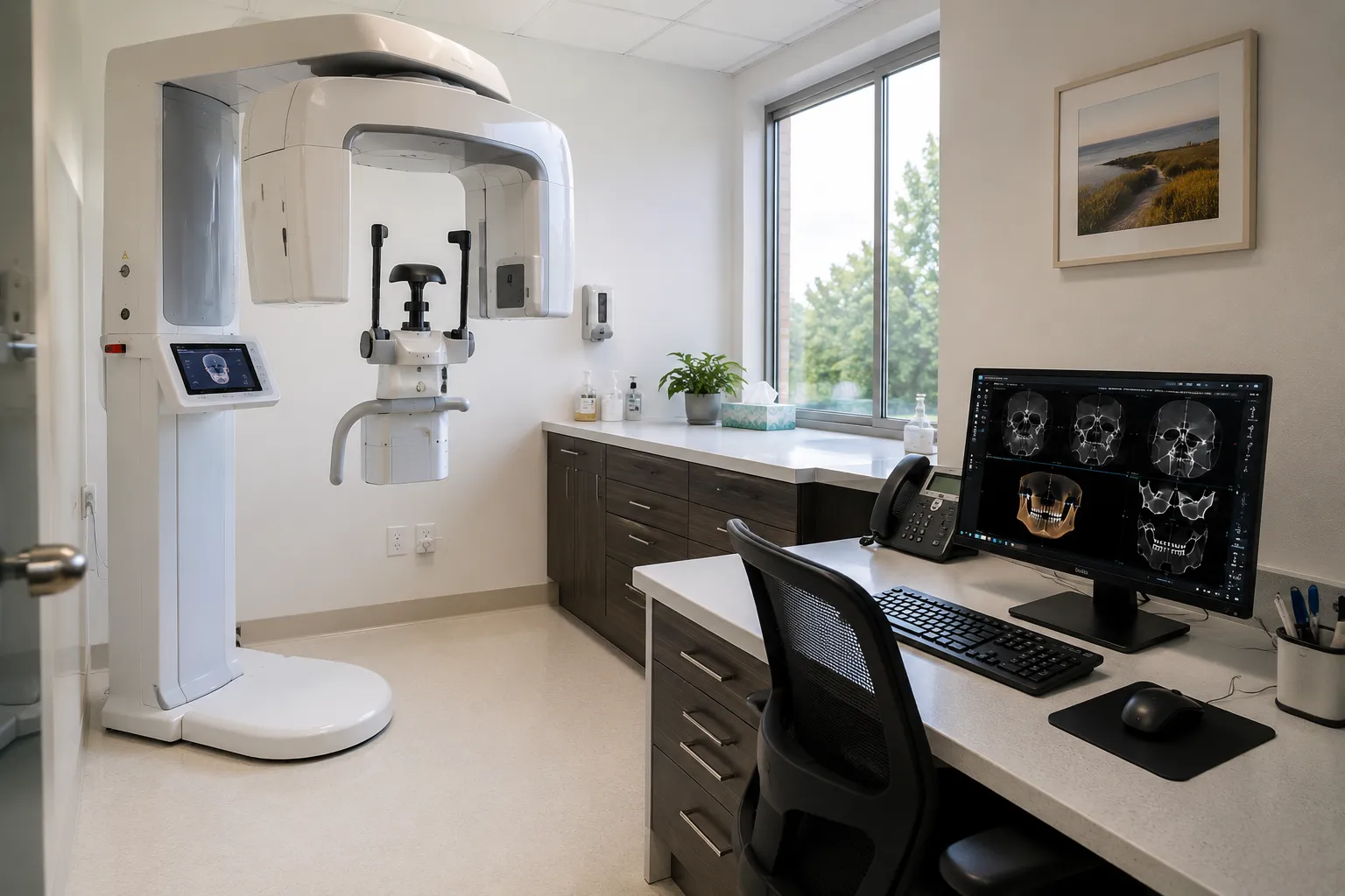
Cone beam computed tomography (CBCT) produces three-dimensional images of the jaw and surrounding structures, supporting implant planning, surgical assessment and other work. For a practice, it is a significant installation involving radiation considerations, room requirements and software, so the decision reaches beyond image quality to how the scanner is justified, sited and supported within a practice's imaging.
The ten scanner types below reflect the range and the considerations a practice weighs. Each entry explains a factor and what to confirm before purchase, with radiation and justification treated as central.
Field of view, dose and clinical need
Field of view determines how much anatomy a scan captures, and the appropriate field follows the clinical need rather than the largest available. Selecting a field of view matched to the examination supports dose considerations, since capturing more than needed is not in a patient's interest.
Dose management is central to any imaging using ionising radiation. Features supporting dose management, and the principle of using the smallest appropriate field of view and settings, matter for every examination, and a radiation protection adviser is involved in the arrangements from the start.
The ten scanner types
1. Small field of view CBCT scanners
Capture a limited region such as a few teeth, suited to focused examinations. Often the appropriate choice for localised assessment.
2. Medium field of view scanners
Capture a larger region such as an arch, supporting implant and surgical planning across a wider area.
3. Large field of view scanners
Capture the jaws and surrounding structures, used where the examination genuinely requires the wider view.
4. Combined CBCT and panoramic units
Provide panoramic imaging alongside CBCT in one unit, covering both two and three-dimensional examinations.
5. Combined CBCT, panoramic and cephalometric units
Add cephalometric imaging as well, suiting practices needing the full range of dental radiography.
6. Scanners with selectable field of view
Allow the field of view to be selected per examination, supporting the use of the smallest appropriate volume.
7. Scanners with dose management features
Emphasise features supporting lower-dose examination where clinically appropriate. Confirm the options provided.
8. Scanners with high-resolution modes
Offer higher-resolution modes for detailed examination where required, balanced against dose considerations.
9. Scanners with planning software integration
Integrate with implant or surgical planning software. Confirm the workflow, formats and software support.
10. Scanners with networked image handling
Manage image volumes into the practice's imaging and record systems. Storage and integration need planning.
Room requirements, justification and the practice
A CBCT installation carries requirements beyond the scanner. Radiation protection arrangements, including room shielding assessed by a radiation protection adviser and the controlled area, are established before use, and the space, power and access the scanner needs should be confirmed with the installer. Justification and reporting are central to CBCT practice: each examination is justified on clinical grounds, and the images are reviewed and reported appropriately, sometimes involving wider expertise for the full volume captured. These responsibilities shape how a practice offers CBCT and should be understood and resourced before the scanner is installed, so the imaging is used appropriately, and the radiation arrangements are sound from the first examination.
What to confirm before ordering
-
Field of view. Confirm the fields available match your examinations, favouring the smallest appropriate.
-
Dose features. Establish dose management options and selectable settings.
-
Combined modes. Confirm panoramic or cephalometric imaging if needed.
-
Room and radiation. Verify shielding, controlled area, space and access with a radiation protection adviser.
-
Software. Establish planning integration, formats and support.
-
Reporting. Confirm arrangements for reviewing and reporting the full volume.
Commissioning, training and servicing
Commissioning should include the radiation protection arrangements, acceptance and image quality checks and confirmation of safety features, with a radiation protection adviser involved and records kept.
Training should cover positioning, field of view selection, dose-aware technique, software use and the justification and reporting responsibilities, so the scanner is used appropriately and safely.
For servicing, agree planned maintenance, image quality checks, radiation safety checks, software support and electrical safety testing. Ensure the practice receives device safety alert notices so any field safety notices reach the dental and imaging teams.
Room, dose and radiation arrangements should be settled with a radiation protection adviser first. Practices planning CBCT can raise installation questions with the Medigear.uk enquiry page.
Final thoughts
Approach dental CBCT as an imaging installation with radiation responsibilities, choosing the field of view around clinical need and the smallest appropriate volume. Confirm room, dose and software arrangements with expert input, and resource justification and reporting before installation, so the scanner serves the practice appropriately and safely.
This article provides general procurement guidance for dental practices and healthcare organisations. It is not clinical or radiation protection advice, and it does not replace manufacturer instructions or expert advice on radiation safety.