An electron microscope forms an image with a focused beam of electrons rather than visible light, letting it resolve detail far below what an optical microscope can reach— routinely a few nanometres, and below one nanometre on the best instruments. Because the wavelength of an accelerated electron is thousands of times shorter than that of light, the roughly 200 nm diffraction limit of light microscopy disappears, and viral particles, cell organelles, basement membranes and metal grain boundaries come into view. For a pathology or materials laboratory,e resolving power is the whole point; the practical work is choosing the right type, housing it properly,y and preparing samples to a standard the instrument can exploit.
The two families a buyer will compare are the transmission electron microscope (TEM) and the scanning electron microscope (SEM). They answer different questions, and many sites that need both buy them separately rather than compromising on one.
TEM Versus SEM
Transmission electron microscope
A TEM passes electrons through an ultrathin specimen and forms an image from the electrons that emerge, much as a slide projector shines light through film. It provides the highest resolution— revealing internal ultrastructure such as viral morphology, glomerular basement membranes in renal biopsy, mitochondria, and cilia. Specimens must be extremely thin, typically 50–100 nm, so that electrons can pass without excessive scattering. Accelerating voltages are high, commonly 80–120 kV for biological work and 200 kV or more for materials science.
Scanning electron microscope
An SEM rasters a fine electron probe across the surface and builds an image from secondary and backscattered electrons emitted point by point, giving a three-dimensional-looking view of surface topography at magnifications from tens to several hundred thousand times. It suits surface morphology, particulates, fractures and device inspection. Lower accelerating voltages, often 1–30 kV, reduce beam damage and charging on delicate or non-conductive samples.
Electron Sources and Column
The electron gun determines brightness, coherence and running cost. A tungsten hairpin filament is cheap and forgiving but less bright. A lanthanum hexaboride (LaB6) source is brighter and longer-lived. A field-emission gun (FEG) provides the finest, most coherent probe and best resolution. It is the choice for high-resolution SEM and analytical TEM, but it has a higher capital cost and stricter vacuum requirements. Electromagnetic lenses focus the beam, and the whole column runs under high vacuum so air molecules do not scatter electrons.
Sample Preparation
Image quality depends as much on bench work as on console operation. Biological TEM work involves chemical fixation (commonly glutaraldehyde then osmium tetroxide), dehydration through graded solvents, embedding in epoxy resin, and cutting ultrathin sections on an ultramicrotome with a glass or diamond knife, followed by heavy-metal staining with uranyl acetate and lead citrate to add contrast. SEM samples are usually fixed, dried — often by critical-point drying to avoid collapse — mounted on stubs, and sputter-coated with a few nanometres of gold, gold-palladium or carbon to make them conductive and stop charging. Preparation ties EM into the wider histology workflow, so it is worth reviewing your buyer requirements for knives, coaters and consumables at the same time.
Analytical Add-Ons
Most modern instruments do more than image. Energy-dispersive X-ray spectroscopy (EDS or EDX) reads the characteristic X-rays a sample emits under the beam to identify and map the elements present, which is invaluable for particulate identification, mineral analysis and locating heavy elements in tissue. TEM systems may add electron diffraction for crystallography, scanning-transmission (STEM) detectors, and electron energy-loss spectroscopy. Decide up front which analytical ports you need, because retrofitting later is costly.
Siting and Environment
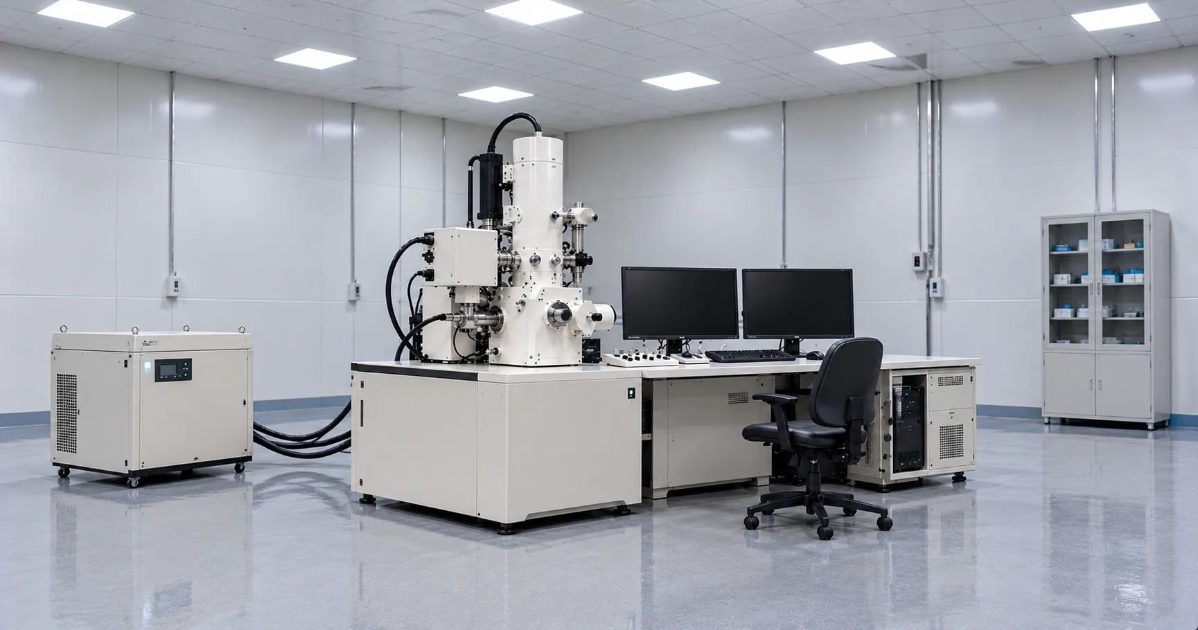
An electron microscope is as much a building project as a purchase. The column is exquisitely sensitive to floor vibration, acoustic noise, stray alternating magnetic fields from lifts and cabling, and temperature drift. High-resolution rooms may need vibration-isolated slabs, magnetic-field cancellation, tight temperature stability and low acoustic noise. Many instruments need a chilled-water supply or a dedicated recirculating chiller, clean single-phase or three-phase power, and space for the vacuum pumps and electronics. Commission a site survey before you commit, because a poor room will cap resolution no matter how good the column is.
Standards, Regulation and Safety
Electron microscopes are laboratory instruments and should carry UKCA or CE marking with electrical safety to the IEC 61010-1 family for measurement, control and laboratory equipment. They generate X-rays internally, so the housing is shielded, and the unit is treated as radiation-safe by design; local rules under the Ionising Radiations Regulations may still apply to registration and monitoring. Sample-preparation chemicals — osmium tetroxide, uranyl acetate, solvents — carry COSHH and, for uranyl salts, radioactive-substance considerations, so fume extraction and waste routes matter. Guidance on device regulation and workplace safety is available from the MHRA and the Health and Safety Executive.
Variants Worth Knowing
Beyond the classic high-vacuum instruments, several variants change what a laboratory can examine. Environmental and variable-pressure SEM tolerate a small gas pressure in the chamber, letting moist, non-conductive,e or uncoated samples be imaged without full drying or coating; useful where coating would obscure the surface. Cryo-electron microscopy holds specimens frozen-hydrated at liquid-nitrogen or liquid-helium temperatures, preserving structure close to the native state and underpinning much modern structural biology, at the cost of a cryo-preparation and transfer chain. Desktop or benchtop SEMs trade ultimate resolution for a small footprint, lower siting demands and simple operation, and suit routine inspection and teaching where nanometre performance is not required. Which variant fits your samples shapes both the specification and the budget.
Detectors and Image Formation
What you see depends on which emitted signal is collected. In SEM, an Everhart-Thornley secondary-electron detector captures low-energy secondaries from the very surface for topographic contrast. In contrast, a backscattered-electron detector reads higher-energy electrons whose yield rises with atomic number, giving compositional contrast that highlights heavy elements. In-lens detectors improve resolution at low voltage. In TEM, the image is captured on a fluorescent screen and, on modern instruments, a direct-detection or CCD camera. Matching detectors to the contrast you need topography, composition,n or internal structure is part of specifying the instrument, not an afterthought.
Procurement Checklist
Before committing to an electron microscope, confirm the following:
-
TEM or SEM (or both) matched to whether you need internal ultrastructure or surface topography.
-
Electron source — tungsten, LaB6 or field emission — set by the resolution and brightness your work demands.
-
Accelerating voltage range appropriate to your specimens and to limiting beam damage.
-
Analytical ports such as EDS, diffraction or STEM specified now rather than retrofitted later.
-
Site survey covering vibration, magnetic fields, acoustic noise, temperature stability and chilled water.
-
Sample-preparation chain — ultramicrotome, sputter coater, critical-point dryer, knives and consumables.
-
Service contract, spares and applications support, including gun and detector lifetimes and response times.
-
UKCA or CE marking and compliance with the relevant electrical-safety and radiation rules.
Total Cost of Ownership
The purchase price is only the start. Field-emission sources, detectors and vacuum components have finite lives and are expensive to replace; service contracts on high-end instruments are a significant annual line; and the preparation lab consumes knives, resins, coating targets and cryogens continuously. Room conditioning, chilled water and uninterruptible power add running costs; weigh these over the instrument's working life, and consider whether an in-house EM is justified against outsourcing occasional work. Standardising on one platform simplifies training, service and spares.
Conclusion
An electron microscope trades the convenience of light for resolving power measured in nanometres, with TEM revealing internal ultrastructure and SEM mapping surfaces in fine relief. Choose the type and source on the questions you need answered, plan the room as carefully as the column, and budget for preparation and service across the instrument's life. To compare electron microscopes, preparation equipment and service options, or to scope a siting survey, talk to the MediGear team.
Disclaimer
This article is for informational purposes only. It is published by MediGear (medigear.uk) for general information and procurement guidance. It is not clinical, diagnostic, treatment, technical, engineering, legal or regulatory advice, nor a product endorsement, guarantee or substitute for professional assessment. MediGear does not provide medical consultations. Buyers should consult their clinical, biomedical, estates, and regulatory contacts and the manufacturer's documentation, and independently verify all specifications, certifications, compatibility, and suitability before purchase. Specifications, certifications and availability are correct at the time of publication and may change without notice. MediGear is a medical-equipment distributor and does not sell medicines or pharmaceutical products.