Lithotripsy is a technique that breaks kidney stones into fragments small enough to pass naturally, and in its best-known form, extracorporeal shockwave lithotripsy, it does so without any incision. A machine generates focused acoustic shockwaves outside the body, aims them precisely using imaging, and delivers repeated pulses that fracture the stone. The fragments then pass out through the urinary tract over the following days. Because nothing enters the body, ESWL remains a mainstay for suitable stones.
For a urology or estates procurement lead, an ESWL system is a significant capital purchase, whether a fixed installation or a mobile shared unit, with imaging, servicing and consumable considerations. This guide explains how it works and what to weigh when buying or contracting for one.
How extracorporeal shockwave lithotripsy works
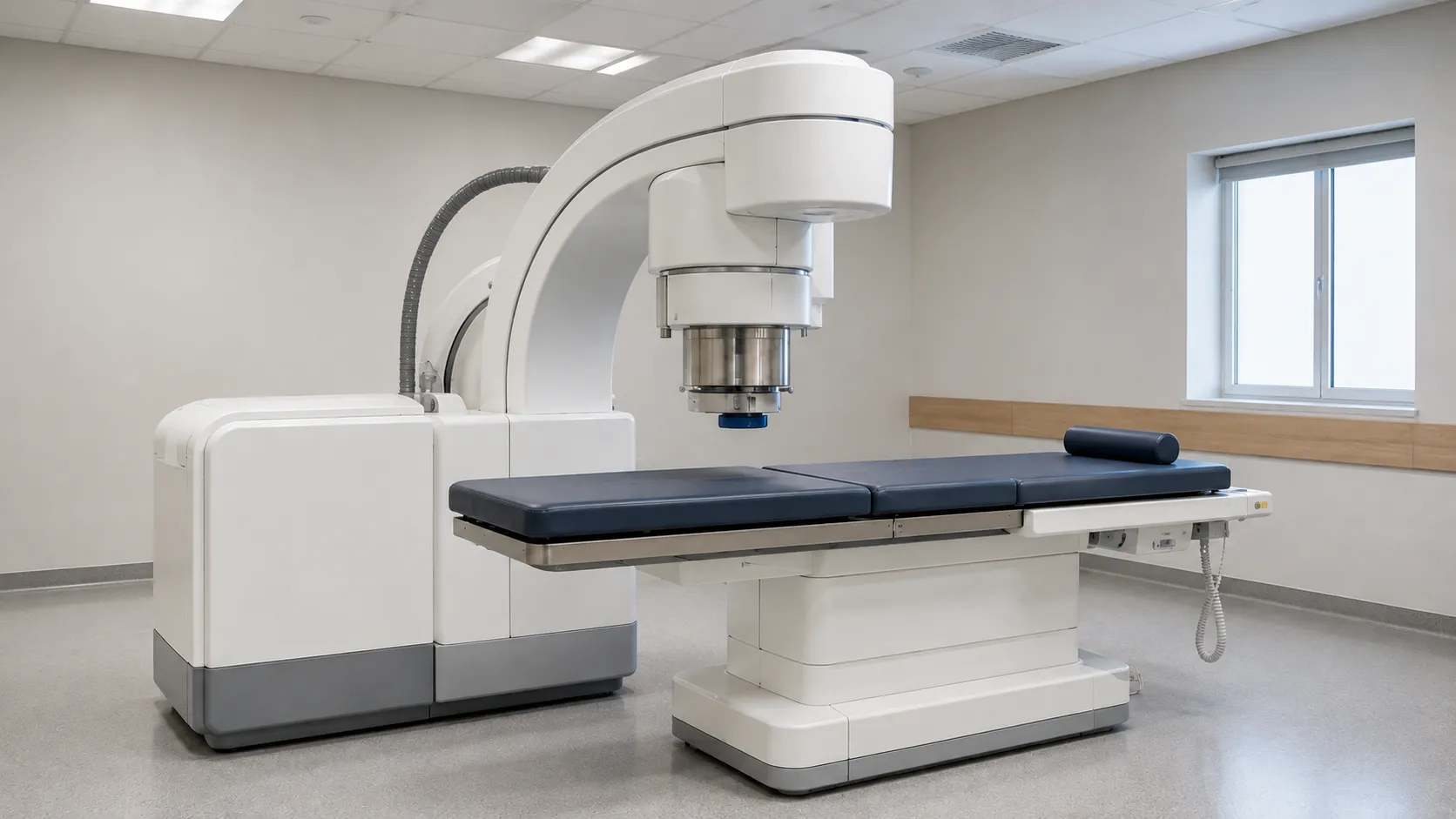
Three elements define the machine: a shockwave source, a coupling to the body, and an imaging system for localisation and targeting. Shockwave is a high-energy acoustic pulse generated and focused so its energy converges at a single point, the focal zone. The patient is positioned so the stone sits at that focal point. Each shockwave passes harmlessly through soft tissue, but at the gemstone, the sudden pressure change and the stress mechanically fracture the crystal structure. Thousands of pulses over a session progressively break the stone into sand-like fragments. The waves are usually delivered in time with the patient's heart rhythm or at a controlled rate to balance effectiveness against tissue effects.
How the shockwave is generated
Three generator technologies are in common use, and the type affects the focal zone, maintenance and consumables:
-
Electrohydraulic — a spark discharge under water creates a shockwave reflected to a focal point. It provides a focal zone,, but thespark electrode is a wearing partpart.
-
Electromagnetic — a coil drives a membrane to generate the pulse, focused by a lens or reflector. It is stable and long-lived, a common choice in modern systems.
-
Piezoelectric — many small piezo elements fire together on a curved surface, focusing energy tightly with a small focal zone and low skin discomfort.
The energy must reach the body without an air gap, so a coupling medium, a water cushion with gel, transmits the waves from the source into the patient. Good coupling, with no trapped air bubbles, is essential to effective treatment.
Imaging and localisation
Accurate targeting is what makes ESWL work, and imaging is central. Systems use fluoroscopy (X-ray), ultrasound, or both to place the stone at the focal point and confirm it stays there. Fluoroscopy shows radiopaque material and is widely used, but it uses ionising radiation, so systems and staff must manage dose. Ultrasound avoids radiation, shows radiolucent stones, allows continuous monitoring, and is valuable for reducing exposure, particularly in younger patients. A dual-imaging machine offers flexibility. Buyers should weigh imaging type against case mix, radiation-protection duties, and the team's skill set.
Kteam's skill set specifications a buyer must check
Decide first between a fixed installation and a mobile or shared unit, since ESWL is expensive and many services share a machine across sites via a mobile provider. Check the shockwave generator type and focal-zone characteristics, and review the imaging configuration, fluoroscopy, ultrasound or dual, and radiation-protection requirements. Consider patient table and positioning, integration with the imaging, and whether the table supports other urological procedures. Assess throughput, ease of coupling and setup time, as well as consumable setup wear and setups; assess consumable gel and cuswear. Finally, weigh the service contract, uptime guarantees, and shockwave-source replacement costs, which dominate the running costs of these machines.
Where ESWL fits and its limits
ESWL is non-invasive and typically day-case, which is its great advantage, but it is not right for every stone. tyStone density and location determine whether shockwave, an endoscopic laser approach, or percutaneous surgery is appropriate; that clinical decision is guided by bodies such as NICE and sits with the urology team, not procurement. Very hard or large stones may resist shockwave and may require an endoscopic laser technique instead. Understanding these limits matters for buyers because it shapes how much ESWL capacity a service actually needs alongside its endoscopic and laser provision, and whether a shared mobile service is more sensible than owning a machine outright.
Use across settings and service models.
Because of the cost, ESWL provision varies. Large urology centres justify a fixed installation running regular lists. Smaller hospitals and networks often use a mobile ESWL service, where a lorry-mounted or trailer unit visits on scheduled days, spreading the capital cost across sites. Some units combine ESWL with a urology table that also supports endoscopy. The right model depends on stone caseload, geography and the balance between ESWL and endoscopic laser services. Modelling annual case numbers realistically prevents both the under-use of machines and reliance on scarce mobile slots.
Consumables, servicing and total cost of ownership
The headline is the capital or contract cost, but running costs are substantial and vary. Electrohydraulic systems consume spark electrodes; membranes and coupling cushions wear out; coupling gel is used in every case; and the shockwave source has a rated life and is costly to replace. It requires its own servicing, and fluoroscopy and radiation-protection quality assurance; service contracts with uptime guarantees are central, because a machine out of action strangles and a stone list. When comparing options, model the cost per treatment across capital, source replacement, consumables and servicing, not just the purchase price. Our team can help you compare owned versus mobile ESWL and scope a service contract — register as a buyer or contact us.
Regulation and safe use
ESWL systems are active medical devices and, where they use X-ray imaging, fall under the Ionising Radiation (Medical Exposure) at Work Regulations 2000, as they carry a valid UKCA or CE marking with instructions for use. The directive on electrical safety is based on the IEC 60601 family. The MHRA regulates medical devices in the UK; radiation and general guidance sits on GOVUK. Workplace safety falls under the HSE framework. Fluoroscopy-equipped systems require radiation-protection arrangements, quality assurance and trained operators. Procurement ensures that imaging, viewing, and radiation-protection provisions are in place; treatment decisions and delivery rest with the clinical team.
Procurement checklist
-
Decide between a fixed installation and a mobile or shared ESWL service for your caseload.
-
Check the shockwave generator type and its on-one and wear, ring wear
-
Review imaging: fluoroscopy, ultrasound or dual, and radiation-protection duties.
-
Assess the patient table, positioning and any multi-use urology capability.
-
Model consumables: electrodes, membranes, coupling gel and cushions.
-
Weigh the service contract, uptime guarantee and shoand shockwave source placement
-
Confirm UKCA or CE marking, IEC 60601 safety and ionising-radiation arrangements.
Conclusion
Extracorporeal shockwave lithotripsy breaks kidney stones with focused acoustic pulses delivered from outside the body, guided precisely by imaging, so most patients avoid surgery altogether. Buying well means choosing between an owned and a shared machine, matching the generator and imaging to your caseload, and modelling the real cost per treatment across the shockwave source, consumables and servicing. Confirm the imaging, radiation protection, service arrangements, and size the capacity to your actual stone workload. MediGear can help you scope lithotripsy provision and the surrounding urology equipment so your stone service runs reliably and cost-effectively.
Disclaimer
This article is for informational purposes only. It is published by MediGear (medigear.uk) for general information and procurement guidance, and is not . It is not a diagnostic, technical, or treatment guarantee or endorsement, professional treatment, or substitute for assessment consultations. Buyers should consult their clinical, biomedical, estates and regulatory contacts, and the manufacturer's documentation, and independently verify all specifications, certifications, compatibility and suitability before purchase. Specifications, certifications and availability are correct at the time of publication and may change without notice. MediGear is a medical-equipment distributor and does not sell medicines or pharmaceutical products.