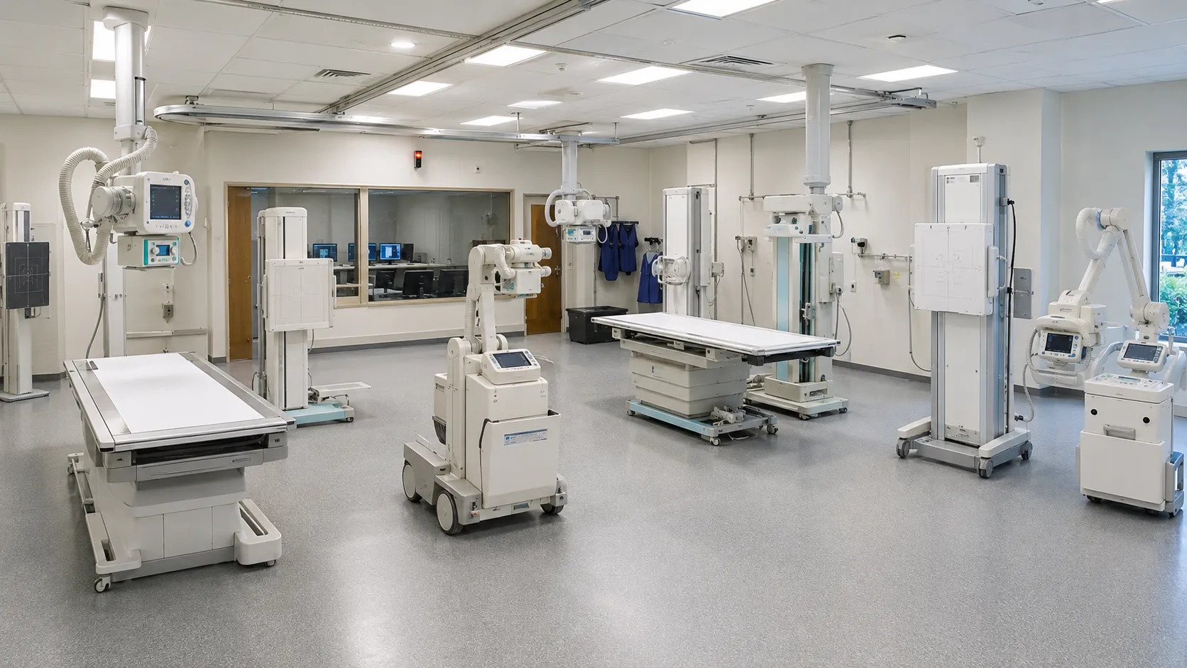
Small imaging units carry a heavy handle caseload with limited rooms, so the system has to be flexible rather than specialised. The decision reaches well beyond the tube and detector: room shielding, floor loading, image transfer and radiation protection arrangements all sit on the same critical path.
The ten system types below cover general radiography, mobile work and theatre imaging. Each entry explains where it fits and what to confirm before ordering.
Settle the room before the system
Work out which room the system will occupy, how it is shielded, where the control area sits and whether the floor and ceiling can take the load. Bring in a radiation protection adviser early, because their input affects layout, shielding and the local rules the unit will work to.
Then decide how images will reach reporting. Detector quality means little if studies stall on their way into the archive, and integration is the element most often underestimated in a smaller unit.
The ten X-ray system types
1. Floor-mounted fixed radiography systems
A column carrying the tube, paired with a table and often a wall stand. Robust and straightforward, they suit general radiography rooms with a standard caseload.
2. Ceiling-suspended tube systems
The tube travels on ceiling rails, providing greater freedom and easier trolley access. Ceiling structure and installation work are the main planning factors.
3. Straight-arm and U-arm systems
Tube and detector held in fixed alignment on a single arm, which speeds positioning and reduces alignment errors. A practical choice for high-volume general radiography.
4. Dedicated chest units
Purpose-built for erect chest imaging, with fast throughput and simple operation. Useful where chest examinations dominate the workload.
5. Mobile radiography units
Wheeled systems taken to the ward, theatre or emergency department. Manoeuvrability, battery endurance and detector handling matter more than maximum output.
6. Battery-powered portable units
Lightweight systems for constrained spaces and outreach work. Confirm the output range against the examination, as portability involves limits.
7. Mobile fluoroscopy C-arms
Used in theatre for orthopaedic, vascular and pain procedures. Check compatibility with your operating tables, particularly the radiolucent area and column position.
8. Mini C-arms
Compact fluoroscopy for extremity work, with a smaller footprint and lower output. They suit hand, wrist and foot procedures rather than general theatre use.
9. Digital radiography retrofit detectors
Portable flat panel detectors that upgrade an existing system without replacing the generator or stand. Confirm software compatibility and detector durability, since panels are handled constantly.
10. Computed radiography systems
Cassette-based imaging plates read by a scanner. Still in service in some units, though support life and plate availability should be confirmed carefully before any further investment.
Specification points that matter
-
Generator output. Match power and exposure range to the examinations performed, including larger patients.
-
Detector type and size. Confirm panel sizes, whether they are wireless, and how they are charged and stored.
-
Image transfer. Establish the connection to your archive and worklist, the standard used, and who configures it.
-
Dose recording. Check how exposure data is recorded and exported for audit purposes.
-
Room requirements. Confirm shielding, electrical supply, floor loading, and any building-required work needed. Ask about expected tube life at your workload and the replacement arrangements.
Commissioning, quality assurance and servicing
Commissioning should include acceptance testing, verification of exposure accuracy, image quality assessment and confirmation that safety interlocks and warning signals work. Agree who performs each test and what documentation the unit receives and retains.
Routine quality assurance then continues at set intervals, alongside staff training in positioning, exposure selection and the local rules for the controlled area. Training should include anyone who operates a mobile unit outside the imaging department.
For servicing, agree planned maintenance, detector cover, tube replacement terms, software support and response times, along with spare parts; the unit receives published device safety alerts and keeps field safety notices visible to the team maintaining the equipment.
Final thoughts
Specify around the room, the caseload and the reporting route. Most small units are well served by one flexible general radiography system, a mobile unit for wards and theatre, and fluoroscopy only where procedures justify it. Settle shielding, image transfer and service cover before delivery, and the unit runs without surprises.
This article provides general procurement guidance for healthcare organisations. It is not clinical, diagnostic or radiation protection advice, and it does not replace manufacturer instructions or expert advice on radiation safety.