Ultrasound buying decisions often focus on the console, when the real difference lies in the probes, the workflow, and the support arrangement. A mid-range system with the right transducers and a sensible reporting path usually outperforms a higher-specified machine that nobody has time to configure.
These ten system types cover the range a general diagnostic clinic is likely to consider, with the practical questions that matter for each.
Define the scanning list before the specification.
Write down what the clinic actually scans and how often. Abdominal, obstetric, musculoskeletal, vascular, small parts and procedural guidance each favour different transducers and settings. The list tells you which probes are essential, which are occasional and which are unnecessary.
Then consider who scans. A clinic with sonographers using the system all day has different ergonomic needs from one where clinicians scan between consultations. Boot time, preset access, and probe changeover speed shape daily use more than headline image-processing claims.
The ten ultrasound system types
1. Cart-based general imaging systems
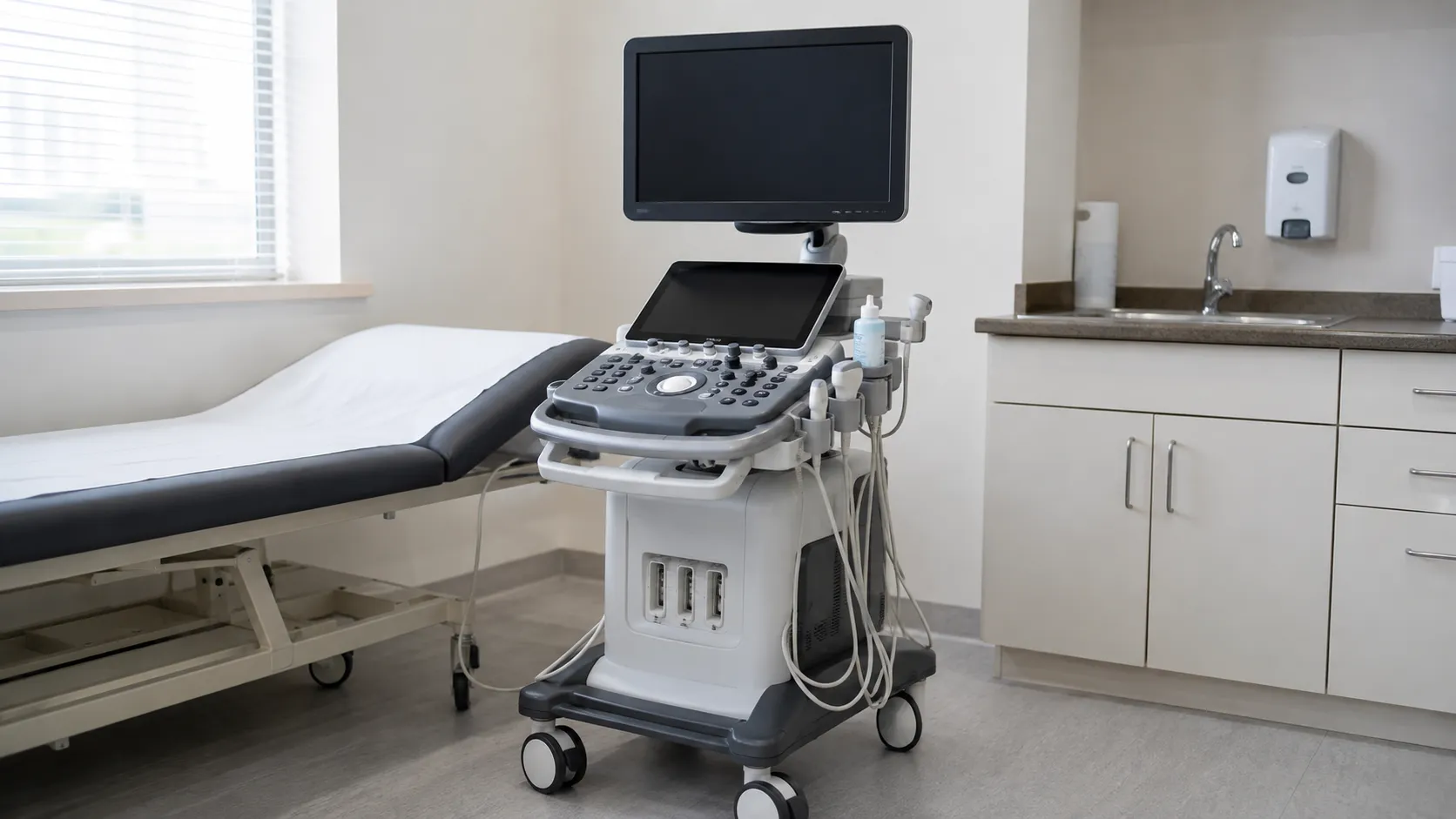
The standard clinic system supports multiple probes, a full preset library, and a proper reporting workflow. Suited to clinics with a varied and steady scanning list.
2. Compact cart systems
Smaller consoles offering most general imaging capability in a reduced footprint. They suit clinics with limited room space or systems that move between rooms.
3. Portable laptop-style systems
Fold-down units carried between sites or clinics. Battery endurance, image storage and connection to the reporting system are the practical questions, not raw processing power.
4. Handheld and probe-based systems
Transducers that pair with a tablet or phone. Useful for bedside assessment and outreach work rather than diagnostic reporting, so define the clinical scope carefully.
5. Systems with colour and spectral Doppler
Doppler capability underpins vascular and much abdominal and obstetric work. Confirm which Doppler modes are included as standard and which are separately licensed options.
6. Musculoskeletal-focused systems
Configured with high-frequency linear probes and dedicated presets for tendon, joint and soft tissue work, often with needle guidance support.
7. Obstetric and gynaecological systems
Set up with curved and endocavity probes, obstetric measurement packages and reporting templates. Confirm which measurement packages come with the system.
8. Systems with three and four dimensional imaging
Volume acquisition and rendering, generally an obstetric or specialist addition. Confirm the probe requirement, as volume imaging usually needs a dedicated transducer.
9. Point-of-care systems
Simplified interfaces built for rapid focused assessment in urgent settings, with quick startup and streamlined presets rather than a full diagnostic feature set.
10. Systems with elastography and advanced applications
Additional tissue assessment tools available on some platforms. Treat these as considered additions where a defined clinical need exists, and confirm licensing and training requirements.
Probes, storage and workflow
-
Probe count and ports. Confirm how many active ports the system has and whether it matches your probe set without unplugging mid-list.
-
Probe availability. Check lead times and replacement supply, since probes are the most commonly damaged component.
-
Image storage. Establish how studies are archived, whether the system connects to your imaging archive, and who configures that link.
-
Reporting. Ask which measurement and reporting packages are included and which are licensed extras.
-
Cleaning. Confirm approved cleaning and disinfection methods for each probe type, particularly endocavity transducers.
Training, servicing and lifecycle
Applications training is the part most often cut and most often regretted. Ask for structured sessions covering preset optimisation, measurement packages, and the reporting workflow, with a follow-up visit some weeks after installation, once real questions have surfaced.
For servicing, agree planned maintenance intervals, electrical safety testing, probe inspection, software update arrangements and response times. Probe repair or replacement terms deserve particular attention because probes are highly exposed to failure due to cable damage and drops.
Confirm expected software support life and spare-parts availability in writing. Registering to receive device safety alerts keeps the clinic informed of field safety notices affecting installed imaging equipment.
Final thoughts
Let the scanning list drive the specification. Most general clinics are best served by a cart-based system with a well-chosen probe set, Doppler as standard, a working link to the imaging archive and applications training booked in twice. Add specialist capability only where a defined clinical need justifies the transducer and the training that comes with it.
This article gives general procurement guidance for healthcare organisations and clinics. It is not clinical or diagnostic advice, and it does not replace manufacturer instructions or professional judgement.