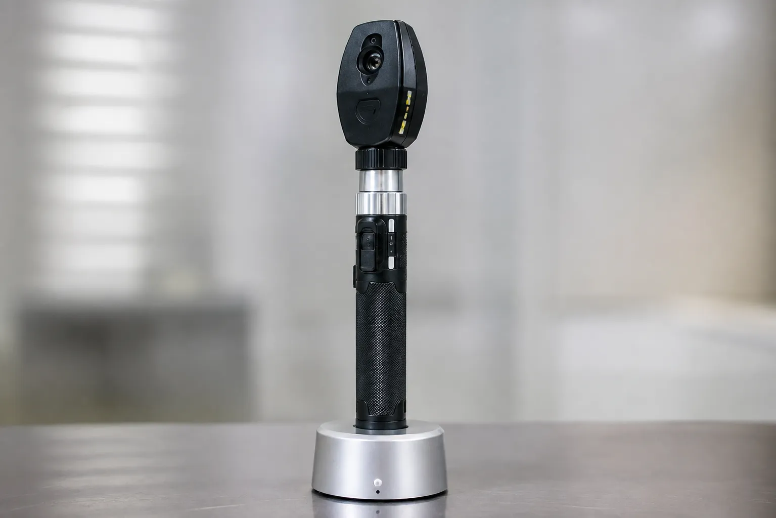
Fundus examination is a skill supported by optics. A poorly lit instrument with a narrow field makes an already demanding examination harder, and it even discourages clinicians from attempting it at all. Choosing well encourages the examination to happen more often, which is usually the point.
The ten instrument types below cover direct examination, wider-field viewing and imaging. Each entry explains its role and what to confirm before purchase.
Optics, illumination and practical use
Three factors shape usability. Field of view determines how much of the retina is visible in one position. Illumination quality determines what is visible through a small or hazy pupil. And working distance determines how close the clinician needs to be, which affects comfort during repeated examinations.
Handle systems matter more than they appear to. A practice standardising on one rechargeable handle platform across ophthalmoscopes and otoscopes reduces spare stock and charging clutter considerably.
The ten instrument types
1. Standard direct ophthalmoscopes
The familiar handheld instrument giving a magnified view of the posterior pole through a narrow field. Widely used in general practice and hospital clinics.
2. Wide-field direct ophthalmoscopes
Optical designs that provide a broader view of the fundus, which many clinicians find easier with an undilated pupil.
3. Ophthalmoscopes with filter and aperture sets
Include red-free, cobalt blue and slit apertures for specific examination tasks. Confirm which apertures are included on the model.
4. Binocular indirect ophthalmoscopes
Head-mounted instruments used with a condensing lens, giving a wide stereoscopic view of the peripheral retina. Specialist training is required.
5. Rechargeable handle systems
Battery handles supporting several instrument heads, charged in a desk or wall unit. Standardising here simplifies both stock and charging.
6. Wall-mounted diagnostic sets
Combined ophthalmoscope and otoscope heads on a wall-mounted transformer with permanent power, suited to clinic rooms with fixed layouts.
7. Digital and video ophthalmoscopes
Capture still or video images to support record-keeping. Confirm image storage and transfer arrangements.
8. Smartphone adapter systems
Attach an optical adapter to a phone camera for image capture. Establish handling and imaging arrangements before adopting them in a clinical service.
9. Non-mydriatic fundus cameras
Table-mounted cameras capturing retinal images that require dilation, used in screening programmes with defined image quality requirements; table handheld fundus cameras
Compact imaging devices for community, domiciliary and outreach screening. Battery life, image quality and transfer method are the practical measures.
Where the examination happens
Room conditions affect fundus examination more than instrument-specific specifications. A room that is well-lit and well-ventilated makes examination easier. A room examination that has a dilated pupil makes it easier. A room that dilates the patient at a comfortable height reduces the strain of repeated examinations across a clinic, where ima. When using, throughout, services are considered; the space needed requires a table-mounted camera and the seating arrangement around it before ordering.
What to confirm before ordering
-
Illumination type. Confirm the light source, expected life and replacement arrangements.
-
Aperture and filter set. Check which options are included rather than assuming a full set.
-
Dioptre range. Verify the correction available range for clinician-patient differences.
-
Handle compatibility. Confirm heads and handles across the practice use one platform where possible.
-
Cleaning. Verify approved cleaning methods for optics and housings.
-
Image handling. For imaging devices, confirm storage, transfer and information governance arrangements.
Training, care and servicing
Instrument care is straightforward but easily neglected. Optics should be cleaned only using the method specified by the manufacturer and stored in a way that protects the lens surfaces between clinics.
Training is worthwhile even for experienced clinicians when a new optical design is introduced, since working distance and pupil handling differ between instruments. For imaging systems, standard image quality is defined as an ungradable image.
For servicing, agree lamp or emitter replacement supply, battery replacement, optical repair arrangements and electrical safety testing for mains-connected sets. Ensure the service receives published device safety alerts so field safety notices reach the clinical team.
Handle standardisation across instrument heads simplifies both spares and charging. Clinics reviewing their diagnostic sets can discuss configurations with the Medigear.uk team.
Final thoughts
Buy the instrument that clinicians will actually use. For most general settings, that means a well-illuminated direct ophthalmoscope with a useful aperture set on a shared rechargeable handle platform, with imaging capability where a screening programme requires documented images.
This article provides general procurement guidance for healthcare organisations and clinics. It is not clinical or diagnostic advice, and it does not replace manufacturer instructions or professional judgement.