
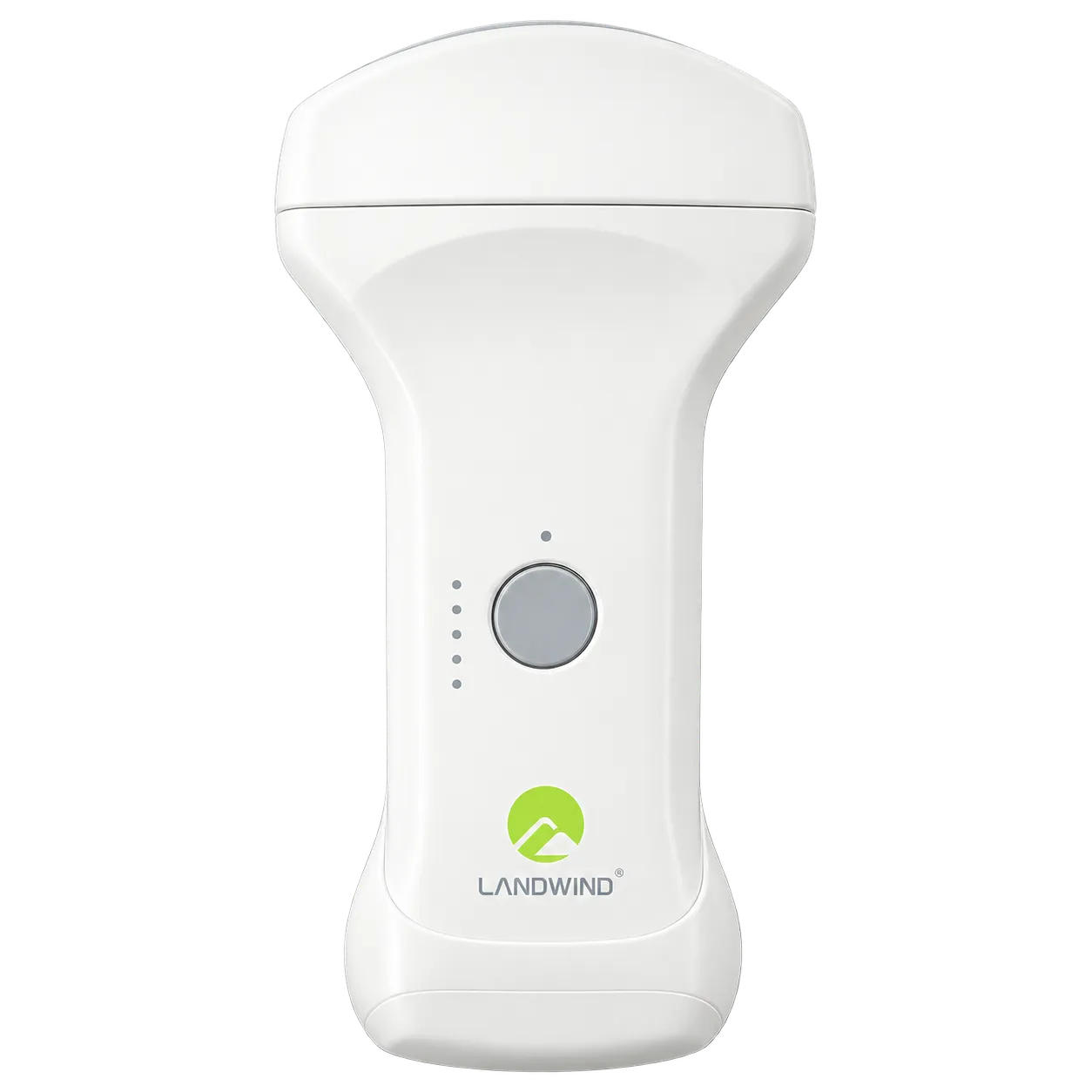
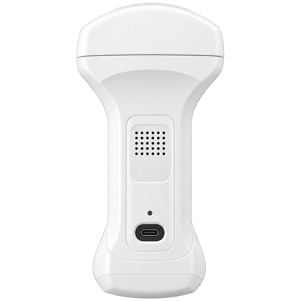
The LX128LC serves clinicians who undertake ultrasound examinations across a range of tissue depths and anatomical regions. Its linear array is designed for superficial structures, where the operating frequencies support imaging of vessels, tendons, small organs, and soft tissue near the skin. Clinicians can use the linear scanning head for vascular assessment, musculoskeletal work, and peripheral nerve imaging. Its shape also suits examination pathways requiring localised probe placement.
The convex end addresses anatomy situated deeper in the body. Abdominal examination is a key use, including imaging of visceral structures and free fluid. The model's declared imaging applications include obstetric, gynaecological, urological, and paediatric scanning. The wider field of view of the curved array supports views of larger internal regions, and the published imaging depth extends to 30 cm. This scanning approach can support general clinical imaging, outpatient departments, and point-of-care assessments.
Cardiac examinations use the cardiac preset associated with the convex transducer. This lets clinicians access a cardiac-focused scanning workflow on the same device used for general and superficial imaging. The precise scanning approach remains dependent on the patient's anatomy and the operator's assessment. M mode adds a time-based view of movement, and optional Doppler modes provide additional flow-focused imaging when available.
At the bedside, the probe's wireless design supports use with a compatible mobile screen or computer. Its compact size suits clinicians who move between rooms and need quick access to different examination types. The scanner also supports freehand guidance during interventions and is cleared for FAST and EFAST assessment.