
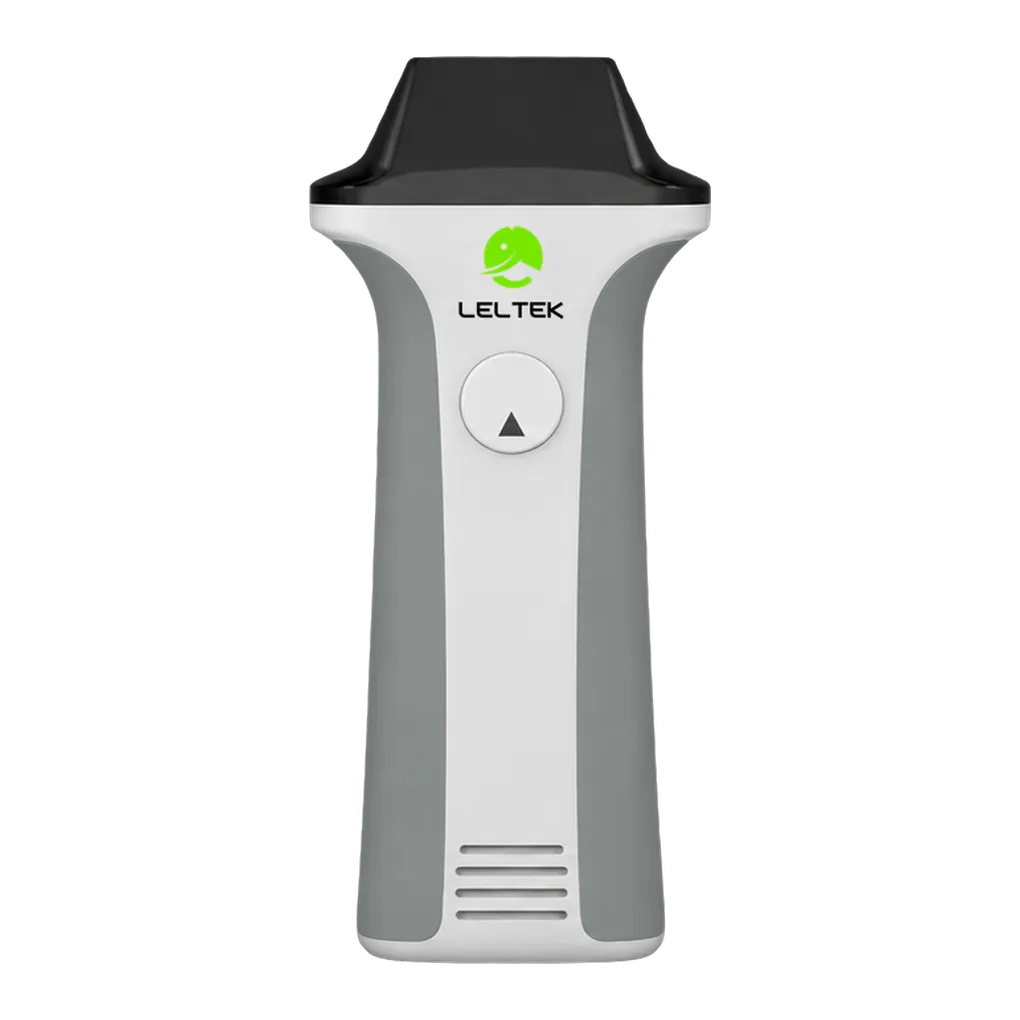
Thyroid and breast examinations suit the LU710LH's focus on superficial anatomy. Its 4.0–17.0 MHz linear array performs the scan, with dedicated presets setting the initial imaging parameters for these organs. B-mode presents tissue structure, and depth and gain controls shape the displayed field. Callipers and annotations add dimensions and examination context to selected frames, creating images you can revisit in the saved patient study.
In musculoskeletal work, the probe supports imaging of conventional and superficial structures, as well as nerve examinations. A live view follows the area beneath the transducer as you move it. The scanner also supports freehand needle and catheter guidance. Freezing a frame creates a measurement point, while cine review retains the surrounding sequence and helps document observations made during the scan.
Peripheral vessel and carotid assessment draw on anatomical imaging and optional Doppler functions. Colour Doppler maps flow within a selected area; power Doppler displays flow signals, and pulsed wave Doppler supports velocity assessment. These views can be recorded alongside the greyscale examination. The app stores images and clips under the patient study, with optional DICOM functions supporting transfer to a compatible clinical archive.
The Aesthetic preset addresses superficial imaging in aesthetic practice, and a dedicated Ocular preset supports ophthalmic examinations. Qualified healthcare professionals use these functions within clinical facilities. Weighing 345 g, the scanner has its own battery and communicates with a compatible phone, tablet, or computer at the examination location. Its diagnostic applications also include paediatric, neonatal cephalic, pulmonary and general abdominal imaging, with recorded frames available for measurement and reporting.