
Peripheral vessel and carotid examinations combine the linear array's anatomical view with optional Doppler assessment. B-mode shows vessel boundaries and surrounding tissue, while colour and power Doppler add flow displays within the selected region. Pulsed wave Doppler provides a spectral view for velocity assessment. A frozen frame can then carry distance markers and annotations into the patient study, linking the measurement with its image.
For thyroid and breast imaging, dedicated presets provide starting settings for superficial anatomy. The 4.2–12.5 MHz bandwidth and adjustable depth support the examination field, with gain controls shaping image brightness. Measurements on frozen images describe distances and outlined areas, while body marks identify the recorded location. Saved frames preserve these observations, along with patient information, for review after the scan.
Musculoskeletal and nerve examinations use live imaging to examine soft-tissue structures as the handheld probe moves across the selected site. The scanner supports conventional and superficial musculoskeletal applications, alongside freehand needle and catheter guidance. Its 200-frame cine buffer holds a sequence for review, and the freeze control pauses a selected view before annotation. This connects live inspection with the measured images retained in the examination record.
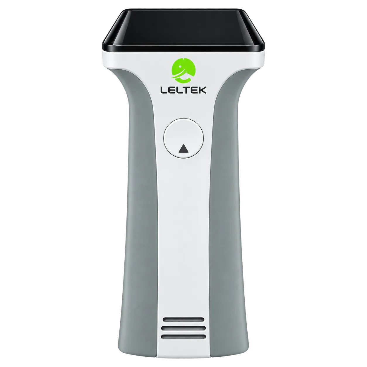
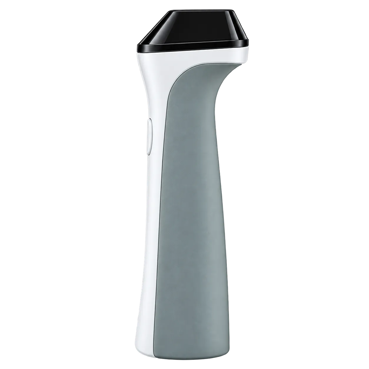
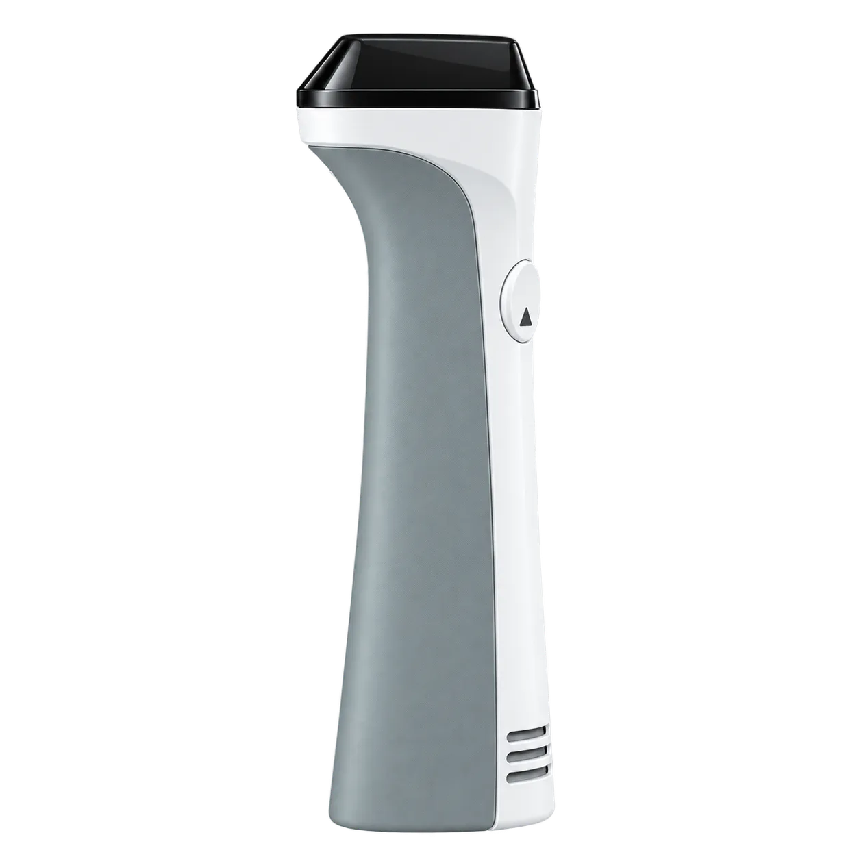
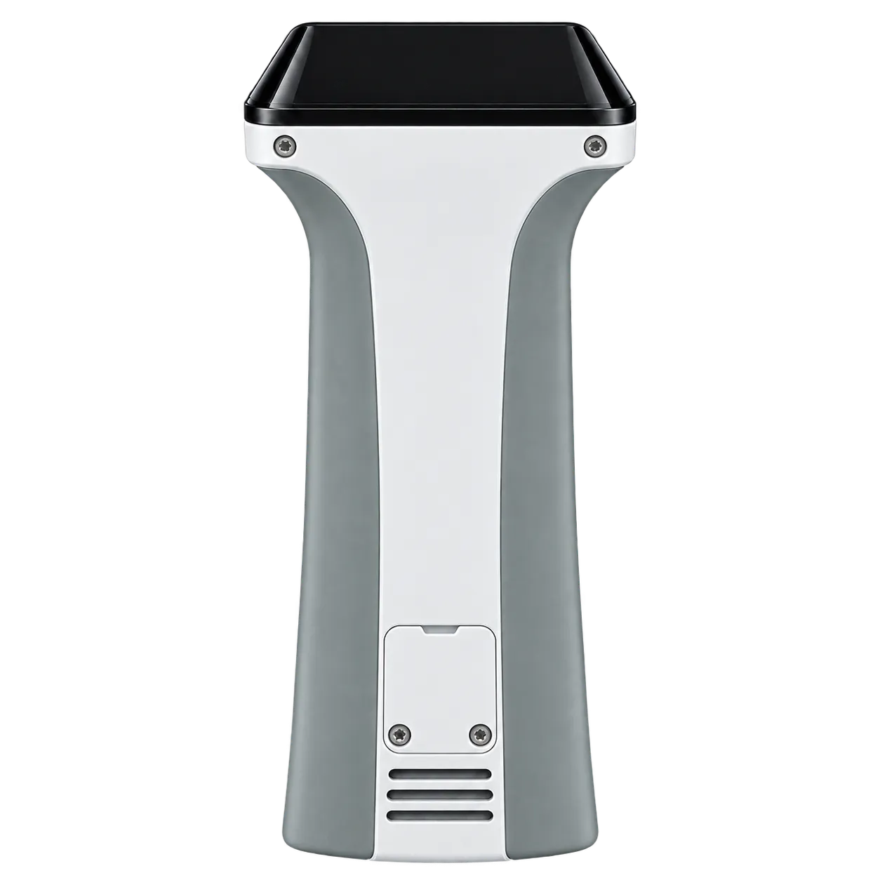
Within hospitals and outpatient facilities, the 357 g probe pairs with a compatible phone, tablet or computer for imaging at the examination location. Additional applications include ocular, paediatric, neonatal cephalic, pulmonary, and general abdominal imaging. Qualified operators select the examination preset and imaging mode through the app. Stills, clips and PDF reports support the resulting record, with optional DICOM transfer linking stored studies to a compatible archive.