
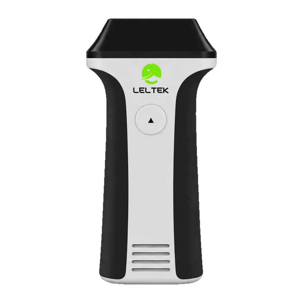
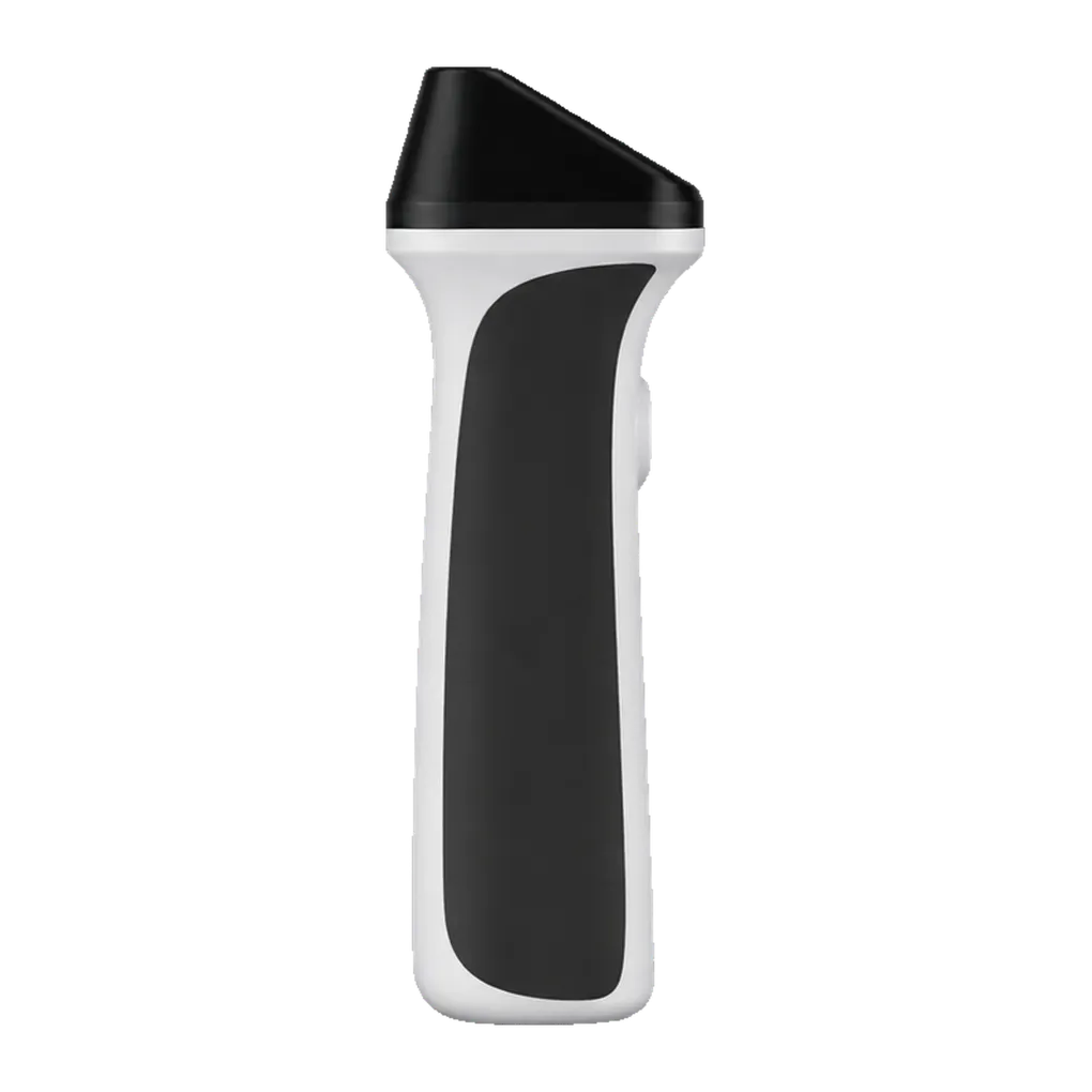
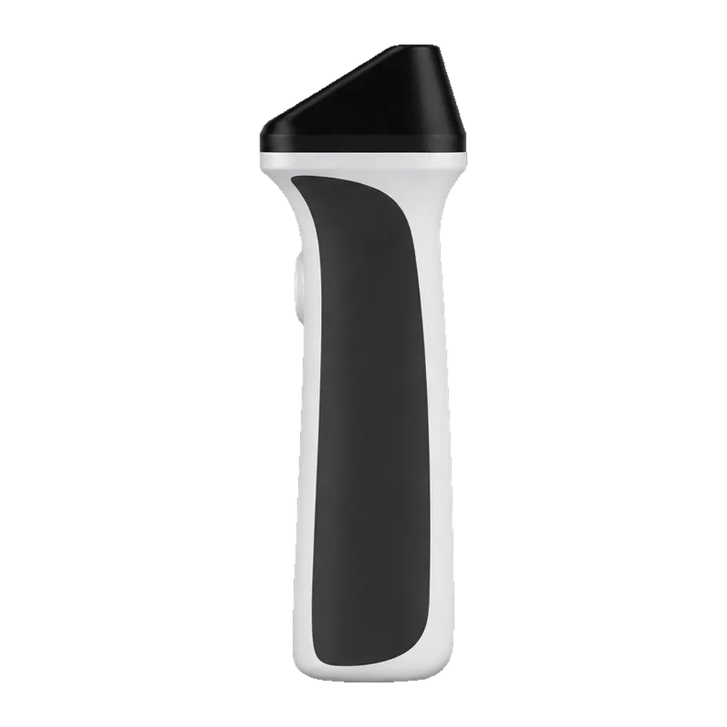
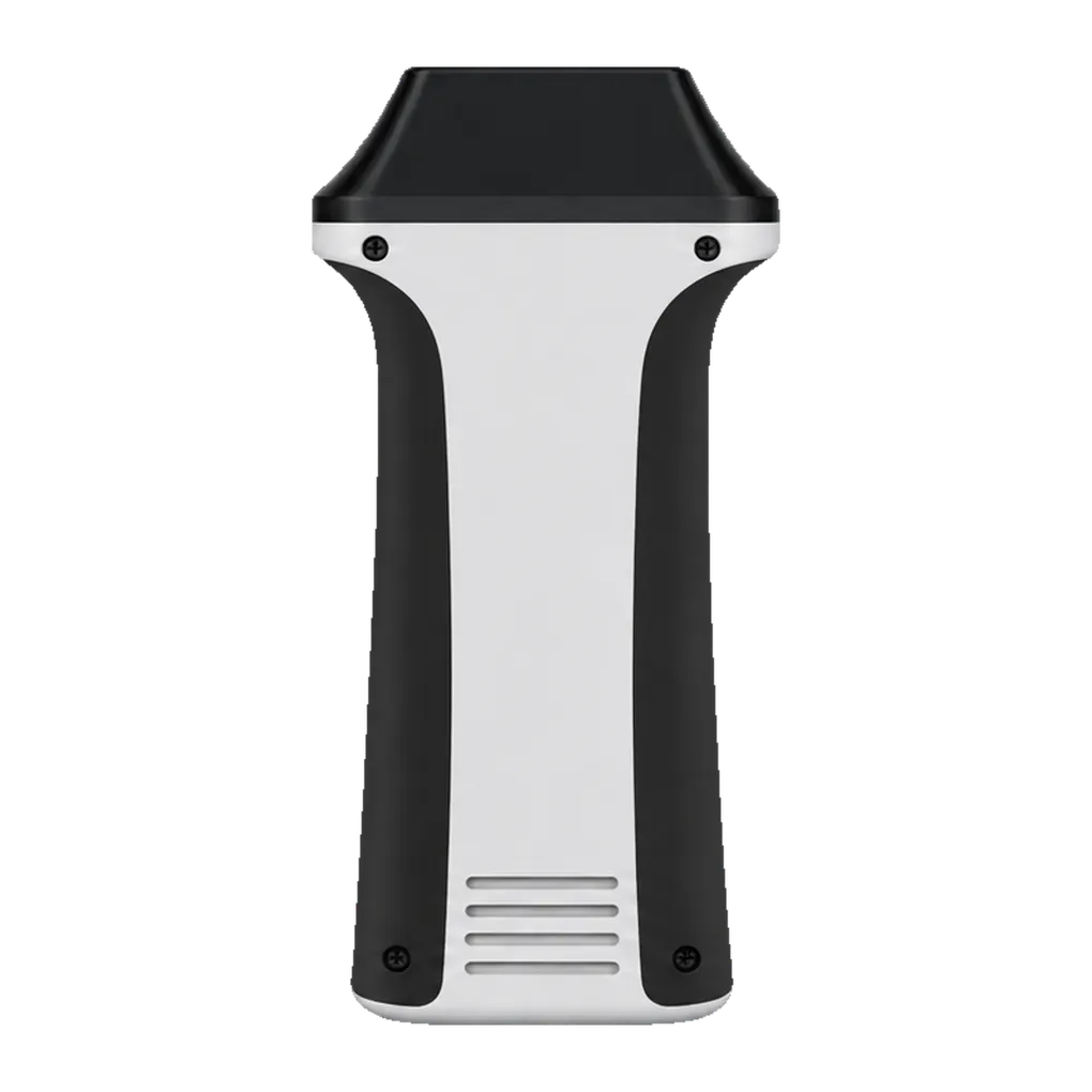
The LU700L serves professional healthcare settings where clinicians examine superficial anatomy or use live imaging to guide freehand needle and catheter placement. Its 5–10 MHz linear array provides imaging depth up to 6 cm. Ultrasound-trained physicians and other qualified healthcare professionals can use that window to view the tissue of interest alongside nearby vessels, nerves and soft structures.
In musculoskeletal clinics, the scanner supports conventional and superficial examinations of muscles, tendons and other soft tissues. The MSK and Superficial presets provide starting configurations for these views. Nerve imaging has its own preset, supporting examinations where the relationship between a nerve and surrounding tissue is part of the assessment.
Vascular work includes peripheral vessels and carotid imaging. Optional colour and power Doppler add blood-flow displays, while optional pulsed wave Doppler provides a velocity trace from the selected sample region. Together, these features extend the information available from a B-mode view during a vascular study.
Small-organ applications include the thyroid, breast, prostate and scrotum within the probe's imaging range. Dedicated Thyroid and Breast presets support two of those examination groups. The indicated scope also includes paediatric, neonatal cephalic, pulmonary and general abdominal imaging where the target anatomy lies within the available 6 cm depth.
The handheld probe can move between examination rooms, procedure rooms, and hospital wards, and it has a manufacturer-compatible display. Saved images, measurements, and annotations retain examination details, while the 200-frame cine buffer supports review of recent scans. Cleaning followed by low-level disinfection forms part of its routine use between intact-skin examinations.