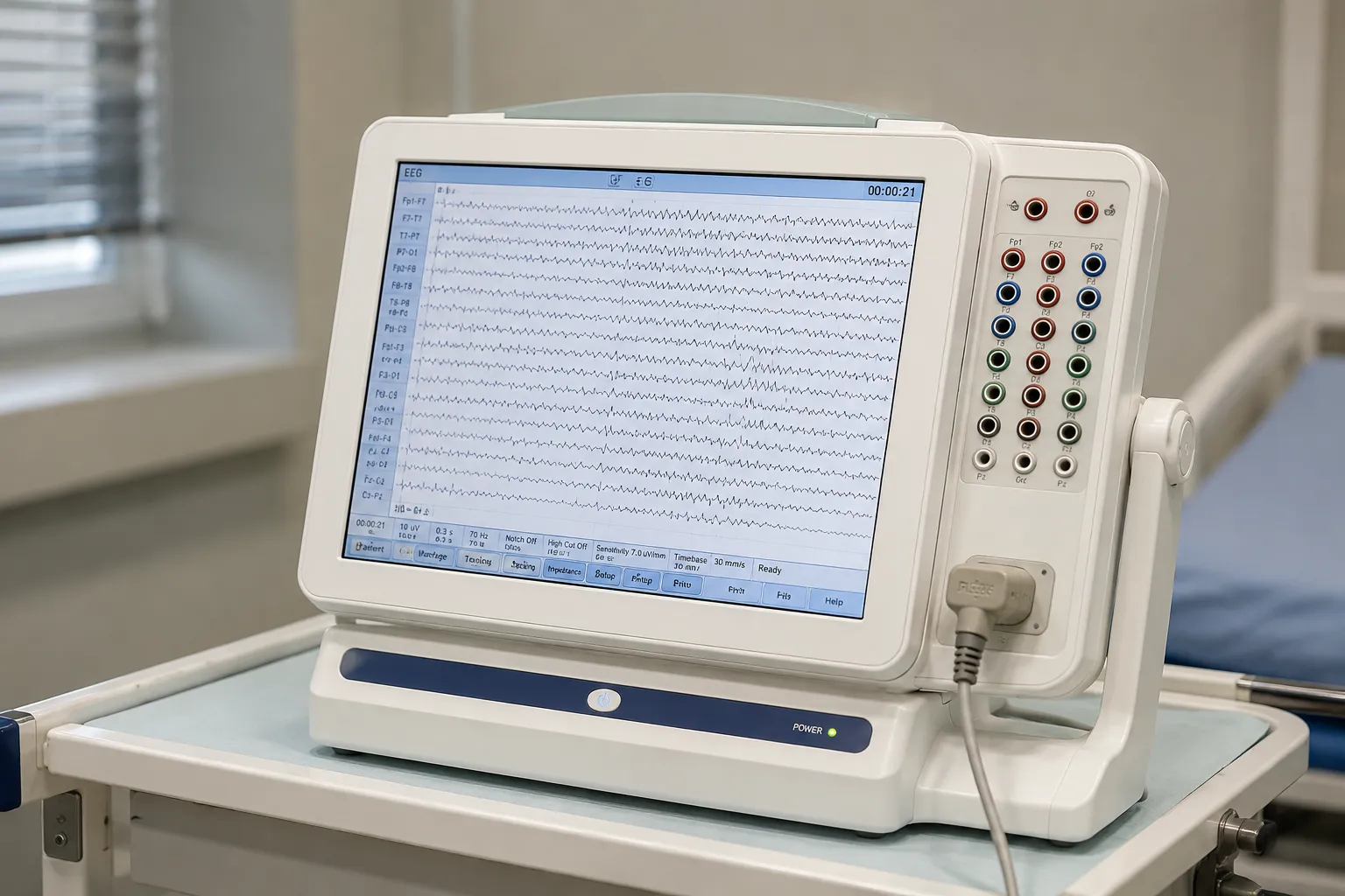
An electroencephalography, or EEG, machine records the brain's electrical activity through electrodes placed on the scalp. It amplifies tiny voltage differences between electrode sites, filters out noise and displays the result as continuous waveforms that a clinician interprets. For a procurement lead, the important questions are channel count, amplifier quality, electrode systems, software, connectivity and the running cost of consumables over the machine's life.
The measurement principle
Populations of neurons in the cortex fire in loose synchrony, producing small fluctuating electrical fields. At the scalp, these signals are in the microvolt range, far smaller than the electrical interference in a typical clinical room. The EEG machine measures the voltage difference between pairs of electrodes, or between each electrode and a reference, then amplifies that difference by a large factor. Differential amplifiers reject signals common to both inputs, which suppresses mains hum and other shared noise. The amplified signal is band-pass filtered, digitised by an analogue-to-digital converter and stored, so the recorded trace reflects genuine cortical rhythms rather than artefact.
How the waveforms are read
Interpreters describe activity by frequency band: delta, theta, alpha, beta and gamma, each spanning a range of cycles per second. They look at amplitude, symmetry between the two hemispheres, the response to eye opening and closing, and reactions to activation procedures such as hyperventilation or photic stimulation with a strobe. Montages, the chosen combinations of electrode pairs, let the reader localise where an abnormal pattern arises. Modern systems display the same recording in several montages and allow re-referencing after acquisition, which is why raw-data storage and flexible review software matter as much as the amplifier itself.
Sub-types and how they compare
Routine and ambulatory EEG
A routine EEG is a short recording in a clinic room. Ambulatory systems are portable recorders the patient wears for a day or more, capturing activity during ordinary life and sleep. Ambulatory units trade some signal control for a far longer sampling window.
Video-EEG and long-term monitoring
Epilepsy monitoring units synchronise EEG with time-locked video so events can be correlated with the trace. These are higher-specification installations with more channels, robust storage and review tools, typically found in tertiary neurology centres.
High-density and intraoperative systems
Research and specialist surgical settings use high-density arrays with many more electrodes for source localisation, or theatre systems for intraoperative monitoring. Most general services do not need this, so avoid paying for channels you will never populate.
Key specifications a buyer must check
Channel count sets what montages you can run; a standard clinical array follows the international 10-20 electrode placement system, so confirm the machine supports at least that with headroom for extra channels such as ECG and EOG. Check the amplifier's input impedance, which should be high, and its common-mode rejection ratio, which should be large, because both determine how well faint signals survive interference. Confirm the sampling rate per channel and the digital converter's resolution. Review the noise floor quoted in microvolts, the available filter settings, and whether the system is electrically isolated to protect the patient. Software matters just as much: re-montaging, spike and seizure detection aids, report generation and secure archiving all affect daily throughput.
Electrodes and consumables
Electrode choice drives both signal quality and running cost. Traditional cup electrodes are applied with conductive paste or gel. They can be reused after cleaning, offering low per-study cost but longer setup. Pre-gelled, disposable electrodes speed application and reduce cross-infection risk but recur as a consumable. Electrode caps with fixed spacing standardise placement and save time on high-channel studies. Whichever you choose, budget for gel, paste, tape, skin-prep and replacement leads, because these small items dominate the ongoing cost of an EEG service.
Standards and UK regulation
An EEG machine is an electrical medical device and must meet the IEC 60601-1 general safety standard, the particular standard IEC 60601-2-26 for electroencephalographs, and IEC 60601-1-2 for electromagnetic compatibility. Patient-applied parts must be suitably isolated. Any device on the UK market needs appropriate UKCA or CE conformity marking and, where required, manufacturer registration. Ask for the declaration of conformity and confirm isolation ratings. The MHRA publishes device safety information and field-safety notices, and clinical professional neurophysiology bodies set standards for recording and reporting, which are equipment vendor.
Cal must follow calibration and decontamination
EEG systems need periodic calibration checks, including a known calibration signal to confirm amplifier gain across channels, plus regular electrical-safety testing of the isolated inputs by your biomedical engineering team. Impedance checks at the start of each recording verify good electrode contact. Reusable electrodes and caps require a validated cleaning and disinfection routine between patients; single-use electrodes remove that step at a consumable cost. Keep leads and connectors on a replacement schedule, because a single damaged lead can corrupt a channel and waste a study.
Care settings and connectivity
EEG is recorded in neurophysiology departments, on wards for bedside and ambulatory studies, in paediatric services with child-friendly caps, and increasingly at home with ambulatory recorders. Veterinary neurology uses similar equipment. Sleep services and epilepsy monitoring units add further demands on channel count and storage. Across all of these, connectivity decides how well the machine fits your infrastructure: confirm networked storage, export in a standard format for sharing between sites, integration with the patient record and remote review so a reporting neurophysiologist need not sit beside the machine. A device that stores raw data openly protects you against being locked into one vendor's review software, and it lets you re-reference and re-montage a recording long after acquisition, which older closed systems prevent. Where studies move between hospitals, an open export format avoids the delay and error of re-recording or manual transcription.
Total cost of ownership, warranty and servicing
The purchase price is a fraction of the lifetime cost. Add electrodes and gels, replacement leads, software licences and updates, calibration, electrical-safety testing and training. Ask suppliers for warranty length, whether software updates are included, typical repair turnaround, loan-unit availability and the cost of spares such as amplifiers and headboxes. The headbox and its leads are the parts handled most and the most likely to need replacing, so confirm their cost early. Standardising on one system across a service simplifies training and stock, and lets studies recorded on one machine be reviewed on another without conversion. A five-year view usually favours a well-supported mid-range system over the cheapest unit whose consumables and downtime erode the saving. The MediGear buyers team can help compare specifications, or you can contact us to check current availability and support options.
Procurement checklist
-
Confirm channel count and support for the 10-20 placement system with spare inputs.
-
Compare amplifier input impedance, common-mode rejection, sampling rate and resolution.
-
Check filter options, noise floor and patient isolation ratings.
-
Decide between reusable and single-use electrodes and price the consumables.
-
Verify UKCA/CE marking, IEC 60601 compliance and the declaration of conformity.
-
Confirm data export, patient-record integration and remote review.
-
Agree calibration, electrical-safety testing and decontamination routines.
-
Get warranty, repair turnaround and spares availability in writing.
Conclusion
An EEG machine turns microvolt cortical signals into traces a clinician can interpret, and the quality of that translation depends on the amplifier, the electrodes and the software behind it. Match channel count and features to your caseload, budget honestly for consumables and safety testing, and confirm standards and servicing before you sign. Comparing verified specifications through MediGear helps you invest in a system your neurophysiology service can rely on for years.
Disclaimer
This article is for informational purposes only. It is published by MediGear (medigear.uk) for general information and procurement guidance and is not clinical, diagnostic, treatment, technical, engineering, legal, or regulatory advice, nor a product endorsement, guarantee, or substitute for professional assessment. MediGear does not provide medical consultations. Buyers should consult their clinical, biomedical, estates and regulatory contacts, and the manufacturer's documentation, and independently verify all specifications, certifications, compatibility and suitability before purchase. Specifications, certifications and availability are correct at the time of publication and may change without notice. MediGear is a medical-equipment distributor and does not sell medicines or pharmaceutical products.