A pachymeter measures corneal thickness in microns, most often at the central cornea. That single figure, the central corneal thickness, changes how eye-pressure results are read and flags corneas that need caution before refractive or cataract surgery. For a buyer, the decision comes down to measurement method, repeatability, workflow fit and the cost of keeping the device running for five years or more.
The measurement principle
Thickness is derived either by sound or by light. Ultrasound pachymetry sends a high-frequency pulse from a probe tip resting on an anaesthetised cornea; the device times the echo returning from the back surface and, knowing the speed of sound in corneal tissue, converts that time into a distance. Optical pachymetry never touches the eye. It measures the offset between reflections from the front and back corneal surfaces, using low-coherence interferometry, Scheimpflug photography or scanning-slit imaging, and calculates thickness from the optical path difference. Because the two methods rely on different assumed velocities and refractive indices, absolute values can differ by a few microns between technologies, which matters when you monitor the same eye over time.
Sub-types and how they compare
Handheld ultrasound units
Compact, battery-powered and comparatively cheap, handheld ultrasound pachymeters are the usual first purchase for a general or optometric clinic. They need topical anaesthetic, a steady hand and disciplined probe hygiene. Readings depend on probe angle, so a tilted contact reads thicker than the true value and operator training becomes a real accuracy factor.
Optical and imaging-based units
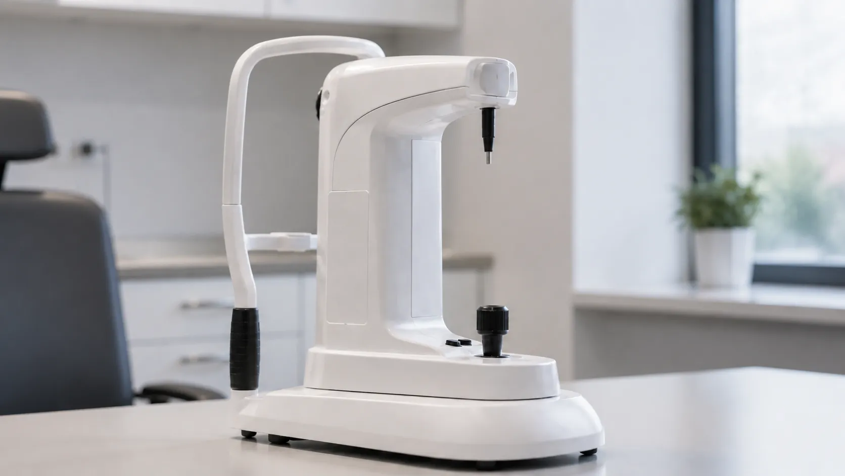
Non-contact optical devices remove the infection-control burden of touching the eye and typically map thickness across the whole cornea rather than a single apical point. They cost more and are frequently anterior-segment tomographers or optical biometers used for surgical planning, so you may already have pachymetry capability within a larger platform.
Specular and confocal variants
Some specular microscopes and confocal systems report thickness alongside endothelial cell data. These are specialist tools for corneal services rather than routine screening. Still, map thickness measurement against your actual caseload before paying for a capability you will rarely use.
Where the readings are used
Corneal thickness feeds glaucoma assessment, because a thin or thick cornea biases tonometry. It underpins refractive-surgery screening, monitors corneal oedema after cataract or graft surgery, and informs some contact-lens fitting decisions. A district general hospital may want mapping tied to its surgical imaging, while a community optometry practice may only need reliable central readings. Match the device to the work, not the widest feature set on the brochure.
Key specifications a buyer must check
Look past the headline accuracy claim. The figure that matters is repeatability, usually quoted as a standard deviation over a set of consecutive readings, because a device that measures the same eye consistently lets you trust change over time. Confirm how many readings the unit averages per measurement and whether it automatically discards outliers, the measurement range, and whether the device supports peripheral points or a full pachymetry map. For ultrasound units, ask the assumed sound velocity and whether it is adjustable. For optical units, confirm the acquisition time, because a slower capture is harder to hold steady on an uncooperative patient. Screen clarity, a clear quality indicator on each capture, and the ability to flag a poor reading all separate a device that is used well from one that is worked around.
Standards and UK regulation
Any pachymeter placed on the UK market must carry appropriate conformity marking, currently UKCA or CE under the recognised transition arrangements, and be registered as required with the regulator. Electrical medical devices should meet the IEC 60601-1 series for general safety, with the relevant IEC 60601-2 particular standard for ophthalmic instruments, and IEC 60601-1-2 for electromagnetic compatibility. Ask the supplier for the declaration of conformity and confirm the device appears in their technical documentation. The MHRA is the reference point for device safety, registration and field-safety notices in the UK, and gov.uk hosts the current guidance on marking and registration timelines.
Calibration and routine maintenance
Ultrasound units are usually checked against a supplied test block or calibration cavity of known thickness; keep that block, log each check and follow the manufacturer's interval. Optical units generally self-check optics at start-up but still need periodic verification and lens cleaning. Whatever the technology, agree who performs calibration, whether it can be done on site or needs return to the vendor, and how downtime is covered. Retain calibration certificates for clinical governance and audit, and retrain operators periodically so technique does not drift.
Decontamination and single-use considerations
Contact ultrasound probes touch the ocular surface, so they need a validated cleaning and low-level disinfection routine between patients, agreed with your infection-control lead. Some services prefer single-use probe covers to simplify reprocessing and reduce cross-infection risk; budget for these as an ongoing consumable. Non-contact optical devices avoid tissue contact entirely, which removes reprocessing but does not replace the need to clean chin rests and forehead bands between patients.
Care settings and portability
Format decides where the device earns its keep. Handheld ultrasound units travel easily between consulting rooms, theatres and outreach or domiciliary clinics, which suits multi-site services and mobile screening. Bench or slit-lamp-mounted optical units stay put, take desk space and reward you with wider mapping and tighter integration with imaging software. Consider who will carry, store and charge a handheld unit day to day, and whether a shared portable device will actually be available when each clinic needs it. Veterinary ophthalmology uses the same handheld ultrasound units so that a portable model can serve a mixed caseload. Environmental factors count too: bright ambient light and an uncooperative patient are easier to manage with a quick contact reading than a longer optical capture, while a c. At the same time, a well-lit room favours richer optical data.
Connectivity and total cost of ownership
A pachymeter only adds value if the reading reaches the record without retyping. Check for export to your electronic patient record, printout options and standard data formats. Then price the whole life of the device: probe tips or covers, disinfectant, calibration blocks, software updates, training and expected repair turnaround. A cheap handheld can cost more over five years than an integrated optical unit if probes wear quickly or downtime is frequent. Ask for a written service plan, warranty length, and spares and loan-unit availability before you commit. If you are equipping several sites, standardising on one model simplifies training and stock. The MediGear buyers team can compare verified specifications, or you can contact us for current availability.
Procurement checklist
-
Confirm the method fits your setting: contact ultrasound for portability, optical for non-contact mapping.
-
Compare repeatability figures and how many readings are averaged per measurement.
-
Check whether central, peripheral or full-map pachymetry is supported.
-
Verify UKCA/CE marking, registration and the declaration of conformity.
-
Confirm calibration method, interval and whether a test block is supplied.
-
Agree probe decontamination or single-use cover costs with infection control.
-
Review data export, patient-record integration and printout options.
-
Get warranty, service turnaround and spares availability in writing.
Conclusion
A pachymeter is a small instrument with an outsized influence on how other eye measurements are trusted. Choose the method that fits your caseload, budget for consumables, calibration and decontamination, honesty, rm sRMety, standards and servicing in writing. Comparing verified specifications through MediGear helps you buy the right unit once rather than replacing it twice.
Disclaimer
This article is for informational purposes only. It is published by MediGear (medigear.uk) for general information and procurement guidance and is not medical, diagnostic, treatment, technical, engineering, legal, or regulatory advice, nor a product endorsement, guarantee, or substitute for professional assessment. MediGear does not provide medical consultations. Buyers should consult their clinical, biomedical, and regulatory contacts and the manufacturer's documentation, and independently verify all specifications, certifications, compatibility, and suitability before purchase. Specifications, certifications and availability are correct at the time of publication and may change without notice. MediGear is a medical-equipment distributor and does not sell medicines or pharmaceutical products.