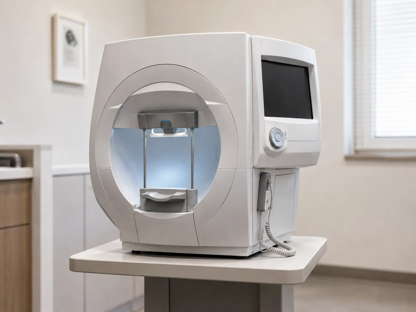
A visual field analyser, also called a perimeter, measures how sensitive a patient's vision is across the whole field of view rather than just what they are staring at. In glaucoma work, it maps areas of reduced sensitivity that signal optic nerve damage, often before the patient notices any loss. For a procurement lead, the analyser anchors an ophthalmology or optometry clinic, so the model you choose shapes throughput, data quality, and years of service cost.
The core answer is simple. The patient fixes on a central target while the instrument presents light stimuli of varying brightness at set positions. The patient presses a button each time they see a stimulus, and the software builds a sensitivity map. Missing points cluster into recognisable patterns that clinicians read alongside optic disc imaging and intraocular pressure.
The Measurement Principle in Detail
Most analysers used for glaucoma run standard automated perimetry inside a bowl of uniform background luminance. A staircase algorithm raises and lowers stimulus brightness at each location to find the dimmest light the eye can detect there, expressed in decibels. That threshold at every tested point becomes the field map. Because sensitivity is measured against a fixed background, the luminance and stimulus calibration must be stable for results to be comparable between visits and between machines.
Threshold Strategies
Older full-threshold routines are thorough but slow, and fatigue itself introduces errors. Faster estimating strategies model the likely sensitivity and test fewer points, cutting test time to a few minutes per eye while holding repeatability. When comparing models, ask which strategies ship as standard and which sit behind a paid software tier.
Test Patterns
Glaucoma testing usually uses a central 24- or 30-degree field. A flexible analyser supports several grids without extra modules, which matters if one machine has to serve a mixed case list.
Perimetry Types and How They Compare
White-on-white standard automated perimetry remains the workhorse for detecting and monitoring glaucoma. Short-wavelength (blue-on-yellow) perimetry and frequency-doubling technology aim to reveal certain damage earlier, but each has trade-offs in noise and patient variability. Kinetic perimetry, where a moving stimulus maps the boundary of the seen field, still has a place in neurological and medico-legal work. Static automated testing dominates glaucoma clinics because it quantifies sensitivity point by point and feeds progression software. If your clinicians want the alternatives, confirm the analyser offers them rather than assuming every unit does.
Key Specifications to Check
Two analysers can look alike and behave very differently in a busy clinic. Weigh the points below against your real case mix and staffing before you commit.
-
Threshold strategies and test patterns included as standard, not chargeable add-ons.
-
Progression analysis software that trends serial fields over years, not just single results.
-
Gaze and fixation monitoring to flag unreliable tests during acquisition.
-
Test duration per eye at typical settings, which drives daily patient throughput.
-
Reliability indices reported, such as false positives, false negatives and fixation losses.
-
Export formats and whether results integrate with your patient record or imaging system.
-
Stimulus source life (bulb or LED) and the cost of trial lenses and other consumables.
-
Documented calibration routine and the interval the manufacturer specifies.
Software, Data and Integration
The hardware captures the field, but the software decides how useful it is. Progression packages that compare a patient's fields across years turn raw sensitivity maps into a monitoring tool, and event or trend analysis is what clinicians rely on to separate real change from test noise. Check whether reports print cleanly, whether data exports in a standard format, and whether the system can sit on your network without breaching information-governance rules. Ask your buyers team to confirm licensing so per-seat fees do not surprise you after installation.
Patient Factors and Reliable Testing
Reliable fields depend as much on the patient and the room as on the analyser. Tired, anxious or first-time patients produce more errors, so a device that keeps tests short and gives clear on-screen coaching pays back in fewer repeats. Room lighting, chair height and a comfortable position all influence fixation, and an unstable head position quietly wrecks a result. When you assess a demonstration unit, watch a real test from the operator's seat rather than the sales slides, and note how the software handles a patient who blinks, drifts or tyres partway through. A machine that flags an unreliable test early, prompts a pause and lets the operator resume without starting again protects both throughput and data quality across a full clinic list.
Standards and Regulation
Any analyser placed on the UK market is a medical device and must carry valid UKCA or CE marking, with the manufacturer meeting obligations overseen by the MHRA. Electrical safety follows the IEC 60601-1 family, and the ophthalmic insts cover general covers and light-hazard requirements for eye instruments. Ask the supplier for the declaration of conformity and confirm the device is registered, because that paperwork is what your governance team needs on file.
Calibration and Routine Maintenance
Perimeters drift. Check stimulus correctness and background luminance; a decibel value means the same thing this year as last. Many units run an internal calibration check, but manufacturers still specify periodic service visits, and bowl-type instruments need the interior kept clean and evenly lit. Build a calibration schedule and a service contract into the decision rather than treating them as an afterthought, and keep dated records so serial fields stay genuinely comparable. A device that logs its calibration status and warns when it is due helps your biomedical engineering team keep the fleet compliant without manual chasing.
Decontamination, Consumables and Care Settings
Patient contact is limited to the chin rest, forehead band, and response button, so decontamination is straightforward: wipe down between patients, following your infection-control policy and the maker's material guidance. Trial lens holders and lenses also need cleaning. Sngs shape the choice: a hospital eye unit may want a fast, high-throughput bowl perimeter with full progression software; a community optometry practice may prefer a compact tabletop unit with a smaller footprint; and screening or domiciliary work leans toward portable perimeters that trade some capability for transportability. Decide where the device will actually work before you fix the specification.
Total Cost of Ownership, Warranty and Spares
The purchase price is only the start. Add software licences, calibration visits, replacement stimulus sources or trial lenses, staff training and eventual disposal. A cheaper unit with costly proprietary consumables can outspend a dearer one within a few years. Weigh how long the manufacturer will support the software and supply spares, because an analyser stranded on unsupported software becomes a liability well before the optics wear out. Check the warranty term, what it covers, response times under any service contract, and whether loan cover exists during repairs. Our supplier partners can help clarify servicing terms before you sign.
Conclusion
A visual field analyser is a long-term clinical asset, so the smart buy balances test speed, reliable progression software, sensible calibration and honest total cost of ownership rather than headline price. Compare models against your real patient flow, verify the UKCA or CE and calibration position, and if you want procurement help sourcing the right unit, contact MediGear to discuss options across our diagnostic equipment range.
Disclaimer
This article is for informational purposes only. MediGear (medigear.uk) publishes it for general information and guidance only and does not provide clinical, technical, or legal advice, or endorse or substitute for professional assessment. MediGear does not provide medical consultations. Buyers should consult their clinical, biomedical, estates and regulatory contacts, and the manufacturer's documentation, and independently verify all specifications, certifications, compatibility and suitability before purchase. Specifications, certifications and availability are correct at the time of publication and may change without notice. MediGear is a medical-equipment distributor and does not sell medicines or pharmaceutical products.