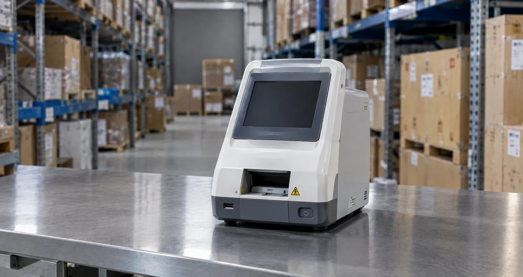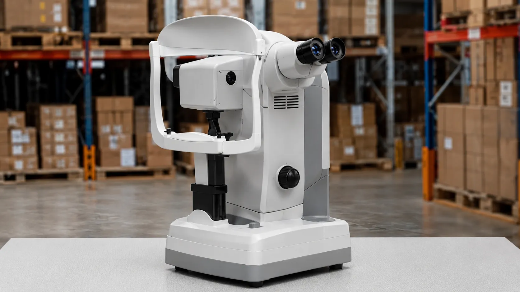
A specular microscope photographs and measures the endothelium, the single innermost layer of cells that lines the back of the cornea. Those cells pump fluid out of the cornea to keep it clear, and they do not regenerate, so counting and assessing them tells a clinician how much reserve the cornea has. The instrument delivers an endothelial cell density figure and a picture of cell size and shape, without touching the eye.
For a buyer, the choice sits between contact and non-contact designs, automated versus manual cell analysis, and how the counts are documented and compared over time. This guide explains the principle, then the specifications and running considerations that matter when you buy one.
The measurement principle
The technique relies on specular reflection. When a narrow beam of light strikes the boundary between the corneal endothelium and the aqueous humour behind it, a small fraction reflects straight back because the two have slightly different refractive indices. The microscope collects that mirror-like reflection at a precise angle and forms a magnified image in which individual endothelial cells appear as a mosaic. Software then identifies cell borders, calculates endothelial cell density (cells per square millimetre, average cell area), the coefficient of variation in cell size (polymorphism), and the percentage of six-sided cells (pleomorphism). Those three shape metrics, alongside the raw count, describe the health of the layer.
Contact versus non-contact designs
Non-contact
Non-contact specular microscopes are by far the most common in clinics. The patient rests at a chin-and-forehead support, fixates a target, and the instrument captures the image across an air gap. There is no touching of the cornea, no anaesthetic drop, and no infection-control contact surface, which makes the examination quick and comfortable, ideal for routine screening.
Contact
Contact instruments apply a sterilised cone gently against the anaesthetised cornea. They can give very high-quality images and work on oedematous corneas where non-contact imaging struggles, but they requ. Still, they require anaesthesia, a disinfection step, and more skill, so they are largely reserved for specialist or research use.
Automated analysis and centre versus corner methods
Older or manual instruments require the operator to mark cells by hand, which is slow and introduces variability. Modern systems auto-capture and auto-analyse, then let the operator correct the cell borders the software has drawn. Two counting conventions exist: the corner method and the centre method, which can yield slightly different densities, so a laboratory should be consistent in which it uses when tracking a patient over time. When comparing instruments, check how the analysis handles low cell counts and guttae, because that is where automated counts can drift.
Why endothelial checks matter for procurement context
Specular microscopy underpins several pathways: assessing donor corneas before transplantation, screening patients before cataract or refractive surgery, monitoring corneal grafts, and following conditions that damage the endothelium. A department's throughput and case mix decide whether it needs a fast non-contact screening tool, a research-grade analyser, or an eye-bank instrument. Knowing which pathway the device serves keeps the specification honest.
Key specifications a buyer must check
Look at the image field and magnification, the automated CAE speed, and the analysis metrics reported—density, cell area, coefficient of variation, and hexagonality. Check how many cells the software analyses per frame and whether it can montage several fields for a larger sample. Assess the fixation and auto-alignment aids that keep a clinic moving, the minimum image quality on hazy corneas, and the ability to image the peripheral cornea as well as the centre. Then weigh the reporting: trend graphs, printouts, and export into your record system.
Repeatability and comparing counts over time
Much of the clinical value comes from watching a cornea trend across visits, so repeatability matters as much as a single accurate reading. A count is only meaningful next to earlier ones if the same region is sampled, the same counting method is applied and the same op,erator technique is used, so consistency of workflow is part of the specification. Ask how the instrument stores prior examinations, whether it displays a trend graph, and how many cells it samples per capture, since a larger sample tightens the density estimate and reduces run-to-run scatter. On corneas with few cells or prominent guttae, automated borders wander, and manual correction becomes essential; a system that makes that correction quick and auditable protects the series' reliability.
Standards, regulation and data handling
A specular microscope is a medical device requiring valid UKCA or CE marking and a declaration of conformity. As mains-powered ophthalmic equipment, it should meet IIEC60601-1 and the relevant particular standards for ophthalmic instruments and light-hazard limits, with IEC 60601-1-2 for electromagnetic compatibility. The MHRA regulates medical devices in the UK and handles safety reporting, with wider guidance on GOV.UK. Because the device stores identifiable images and measurements, confirm its data export and storage meet your information-governance policy, especially where results feed a transplant or surgical pathway.
Calibration, maintenance and decontamination
Non-contact instruments need little between patients beyond cleaning the chin and forehead rests, so single-use tissue covers are the main consumable. Contact instruments require a validated disinfection cycle for the applanating cone and sterile single-use tips or covers where specified. All specular microscopes need periodic verification that the magnification and measurement scale remain accurate, plus routine electrical safety testing. Agree with the supplier on how calibration is confirmed, illumination-source life, and service support terms, because an out-of-calibration density figure undermines every clinical decision built on it.
Consumables, warranty and total cost of ownership
For a non-contact unit, the recurring costs are modest: cleaning materials, chin-rest tissues, occasional lamp or LED replacement, printer supplies, and any software or storage fees. Contact instruments add anaesthetic drops, disinfectant and single-use tips. The bigger numbers are the warranty length, the service contract, and software or analysis-module upgrades over the device's life. Ask about spares availability, loan cover during repair, and whether the analysis software carries ongoing licence costs, then compare on cost per examination over several years rather than the purchase price alone.
Procurement checklist
-
Decide whether you need contact-based or non-contact-based counting, based on your case mix and infection-control preference.
-
Confirm the analysis reports density, cell area, coefficient of variation and hexagonality.
-
Check auto-capture, auto-analysis and manual correction, plus consistency of the counting method.
-
Verify performance on hazy or low-count corneas if you monitor grafts.
-
Request UKCA or CE marking, IEC 60601 evidence and light-hazard compliance.
-
Confirm export and storage meet your governance, especially for transplant pathways.
-
Agree calibration checks, warranty, service, spares and loan cover.
Use across care settings.
Corneal and cataract services use non-contact microscopes for pre-operative screening and graft follow-up. Eye banks need reliable, well-documented counts to assess donor tissue. Refractive clinics screen before surgery. Research units may favour contact instruments for image quality on difficult corneas. Portability is rarely a requirement here, so image quality, analysis accuracy and reporting usually outrank footprint. Match the instrument to the pathway it will serve most.
Conclusion
A specular microscope is judged on the trustworthiness of its cell counts and how easily those counts document a cornea over time. Decide between contact and non-contact, insist on accurate automated analysis with sensible manual correction, confirm the regulatory paperwork and plan for calibration. MediGear can help you compare instruments and arrange servicing so the numbers you rely on stay reliable — register as a buyer or contact our team.
Disclaimer
This article is for informational purposes. MediGear publishes it for general information and legal or regulatory guidance. It is not clinical, engineering, legal, or regulatory advice, nor a product endorsement, guarantee, or substitute for professional assessment. MediGear does not provide medical consultations. Buyers should consult their clinical, biomedical, estates and regulatory contacts, and the manufacturer's documentation, and independently verify all specifications, certifications, compatibility and suitability before purchase. Specifications, certifications and availability are correct at the time of publication and may change without notice. MediGear is a medical-equipment distributor and does not sell medicines or pharmaceutical products.