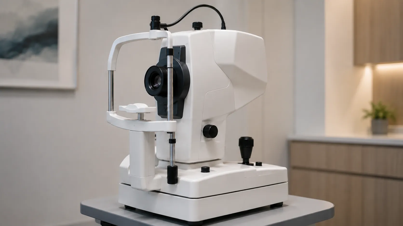
A fundus camera photographs the interior back surface of the eye — the retina, optic disc, macula and retinal vessels — through the pupil. It gives a permanent, shareable record of the fundus that can be compared over time, sent for a second opinion, or fed into a screening pathway. Unlike an examination note, a good fundus image is objective evidence that can be re-graded years later.
For procurement, the decisions are practical: mydriatic or non-mydriatic, tabletop or handheld, field of view, resolution, connectivity to your image-management system, and whether the camera fits the setting where it will actually be used. Get those right, and the camera earns its keep in screening and monitoring alike.
How the image is captured
A fundus camera is essentially a specialised low-power microscope with an attached camera and its own illumination path. Light is directed through the pupil to illuminate the retina, and the sensor captures the reflected image. The result is a flat, evenly lit photograph of a curved surface at the back of the eye. Filters and different light sources extend the technique to fluorescein and indocyanine-green angiography, red-free imaging and autofluorescence, each highlighting different structures.
Mydriatic, non-mydriatic and hybrid
Non-mydriatic
Non-mydriatic cameras use infrared light to focus and align in a darkened room, so the pupil stays naturally dilated and the patient is not given dilating drops. They are fast, comfortable and ideal for diabetic eye screening and high-throughput clinics.
Mydriatic
Mydriatic cameras require pharmacological dilation and generally deliver wider fields and higher-quality angiography, suiting hospital retinal services. Many modern tabletop cameras are hybrid and can switch between modes.
Tabletop versus handheld and widefield
Traditional tabletop cameras give the best image quality and ergonomics for a fixed clinic. Handheld and smartphone-based cameras trade some quality for portability, reaching bedbound patients, care homes, domiciliary visits and community screening. Widefield and ultra-widefield systems capture far more of the peripheral retina in a single shot, useful for screening peripheral disease, though they cost considerably more. Match the form factor to where imaging happens, not to the brochure.
Key specifications a buyer must check
Field of view, quoted in degrees, sets how much retina appears in one frame — 30 to 45 degrees is standard, with widefield reaching much further. Sensor resolution and optical quality determine how small a lesion you can resolve. Check the minimum pupil size the camera can work through, the working distance and auto-alignment features that speed up a clinic, and the imaging modes included, such as colour, red-free and autofluorescence. Assess the software: image capture, patient records, montage stitching, and export in standard formats. Finally, weigh the physical footprint and the table or trolley it needs.
Connectivity and image management
A fundus image is only useful if it reaches the grader and the record. Confirm the camera exports to standard formats and, where relevant, supports DICOM so images flow into your PACS or electronic record. For screening programmes, integration with the grading software and patient administration system is often more important than a marginal difference in image quality. Ask how images are stored, backed up and de-identified for referral, and whether the workflow meets your information-governance requirements.
Image quality and common capture pitfalls
The best sensor cannot rescue a poorly captured image, so a buyer should judge how forgiving a camera is in real hands. Small pupils, media opacities such as cataract, and patient movement all degrade a fundus photograph, producing haze, uneven illumination, or reflective arcs at the edges. Cameras that auto-detect focus and alignment, flag poor-quality frames, and guide the operator to reposition cut the number of unreadable images and the recalls that follow. In screening programmes, an unreadable-image rate translates directly into repeat appointments and cost. Ask to trial the camera on a range of patients, including older eyes and small pupils, rather than relying on brochure images of ideal fundi. A camera that is quick to align and tolerant of imperfect conditions will out-earn a technically superior instrument that only performs on cooperative patients, so weigh ease of capture as heavily as raw resolution when you compare the shortlist.
Standards, regulation and data
A fundus camera is a medical device and must carry valid UKCA or CE marking, backed by a declaration of conformity. As mains-powered electrical equipment with an illumination source directed at the eye, it should comply with IEC 60601-1 and the relevant particular standards for ophthalmic instruments and light-hazard limits, plus IEC 60601-1-2 for electromagnetic compatibility. The MHRA regulates devices in the UK, and NHS diabetic eye screening standards are published through GOV. UK GOV.UK and NHS guidance —UK guidance. Because the camera handles identifiable clinical images, confirm the data-handling arrangements suit your governance policy.
Calibration, maintenance and decontamination
Optical instruments need routine care: keep the lens clean, verify alignment, and perform periodic clinical safety testing. The forehead and chin rests that touch each patient must be cleaned between uses, and single-use tissue covers or wipes are typical consumables. Agree a service schedule with the supplier, including lamp or LED illumination life, sensor and software support, and calibration where the manufacturer specifies it. Handheld units add battery health and physical robustness to the maintenance list.
Consumables, warranty and total cost of ownership
Running costs are modest but real: cleaning materials, chin-rest tissues, occasional illumination-source replacement, software licences and any cloud storage or grading-platform fees. The larger figures are the warranty, the service contract and software support over the device's life. Ask about spares availability, loan cover during repair, and whether software updates and modality add-ons carry ongoing fees. For screening programmes, throughput per session and the cost per patient over several years matter more than the sticker price.
Procurement checklist
-
Decide mydriatic, non-mydriatic or hybrid to match your clinics and patients.
-
Confirm field of view, resolution and minimum pupil size suit your caseload.
-
Check imaging modes — colour, red-free, autofluorescence, angiography — against clinical need.
-
Verify DICOM and export compatibility with your PACS or record system.
-
Request UKCA or CE marking, IEC 60601 evidence and light-hazard compliance.
-
Cost cleaning consumables, illumination-source life and any software or storage fees.
-
Agree warranty, service contract, spares and loan cover.
Use across care settings.
A hospital retinal clinic will want a high-quality mydriatic or hybrid tabletop camera with angiography and widefield options. A community diabetic eye screening service prioritises a fast non-mydriatic camera and tight integration with grading software. GP practices, optometrists and care-home outreach may choose handheld or portable cameras for reach and cost. Veterinary and paediatric use bring their own alignment and handling needs. The right camera is the one that fits the setting and workflow you are equipping.
Conclusion
Buying a fundus camera comes down to matching capture mode, field of view, image quality and connectivity to where and how you image the retina. Confirm the regulatory paperwork, plan for cleaning and servicing, and cost the whole device over its full life rather than the headline price. MediGear can help you compare tabletop, handheld and widefield options and arrange support — register as a buyer or get in touch.
Disclaimer
This article is for informational purposes only. It is published by MediGear (medigear.uk) for general information and procurement guidance and is not clinical, diagnostic, treatment, technical, engineering, or regulatory advice, nor a product endorsement, guarantee, or substitute for professional assessment. MediGear does not provide medical consultations. Buyers should consult their clinical, biomedical, estates and regulatory contacts, and the manufacturer's documentation, and independently verify all specifications, certifications, compatibility and suitability before purchase. Specifications, certifications and availability are correct at the time of publication and may change without notice. MediGear is a medical-equipment distributor and does not sell medicines or pharmaceutical products.