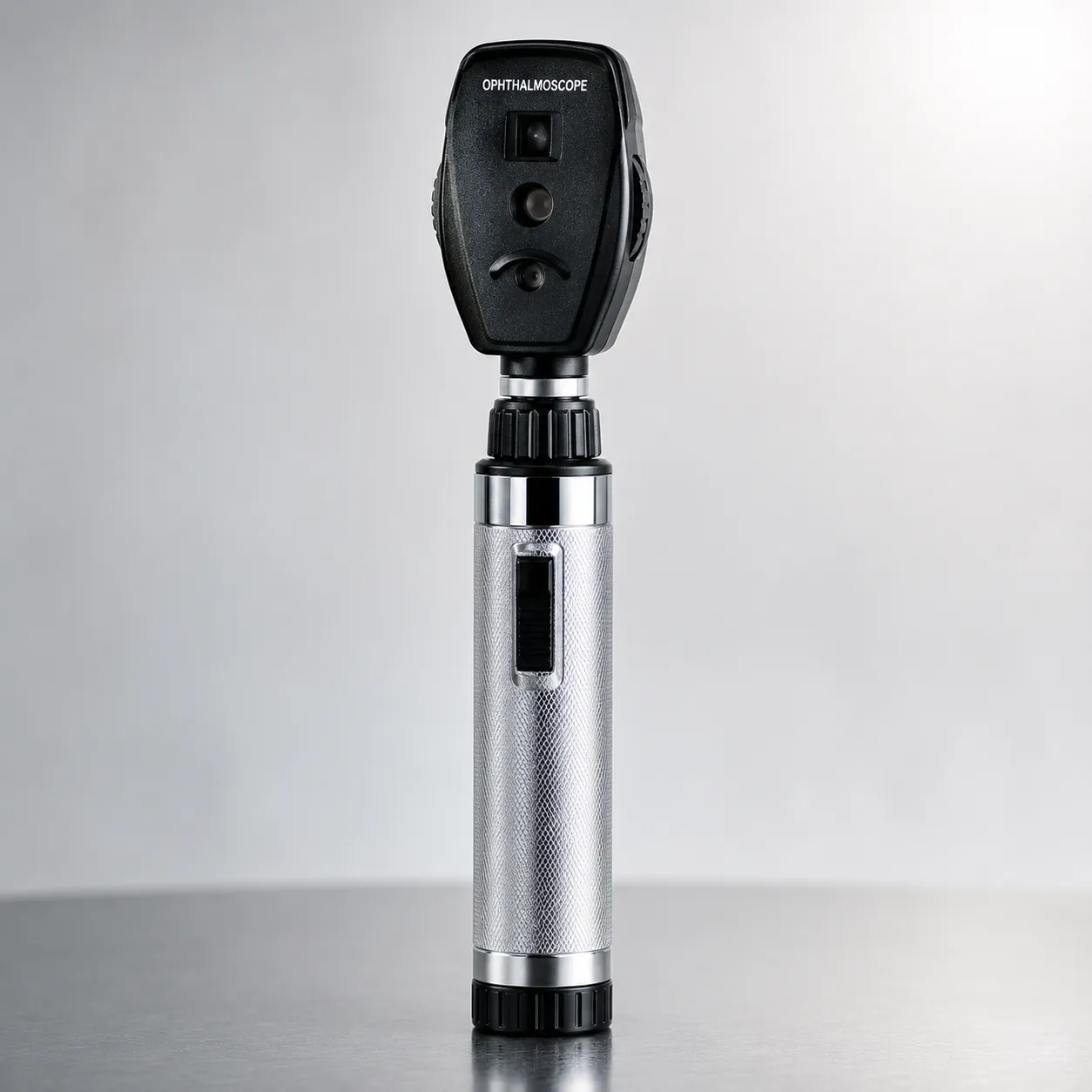
An ophthalmoscope is an optical instrument that lets a clinician look through the pupil to inspect the fundus — the retina, optic disc, macula and retinal blood vessels — at the back of the eye. A light source is directed into the eye, and the examiner views the reflected image, either directly through a handheld head or indirectly through a lens held in front of the eye. Fundus examination supports assessment of the eye and of systemic conditions that leave signs on the retina.
For a buyer, the choice runs from an inexpensive pocket direct ophthalmoscope to a head-worn indirect system and, increasingly, portable and video fundus cameras. This guide explains how ophthalmoscopy works, the main types, the specifications to check, and the standards, maintenance and running costs that shape a good purchase.
How ophthalmoscopy works
The challenge is that the same pupil both admits light and returns the image, so the instrument must illuminate the fundus and let the examiner see it without the two light paths interfering. A direct ophthalmoscope solves this with a compact optical head that gives an upright, highly magnified view of a small field. An indirect ophthalmoscope uses a separate condensing lens held near the patient's eye, producing an inverted, wider-field, stereoscopic view with a brighter light, which is better for examining the peripheral retina. Dilating drops are often used to widen the pupil and improve the view, particularly for indirect examination.
The main types you will compare.e
Direct ophthalmoscope
Handheld, battery or rechargeable, giving a magnified upright image of a narrow field. It is the everyday instrument for general practice, wards and undergraduate teaching — portable and affordable but limited to a small central view.
Indirect ophthalmoscope
Usually head-worn with a separate condensing lens, giving a wider, stereoscopic, brighter view of the retina including the periphery. It is the specialist ophthalmology tool, needing more skill and a hands-and-lens technique.
Wide-field and portable fundus cameras
Wide-field handheld ophthalmoscopes enlarge the field of view over a classic direct head, easing examination through a smaller pupil. Portable and benchtop fundus cameras capture retinal images for documentation, screening and referral, blurring the line between examination and imaging.
Key specifications a buyer must check
Look at illumination type and brightness, colour rendering, and the range of apertures and filters, since a red-free (green) filter aids inspection of blood vessels and the nerve-fibre layer. Check the lens or dioptre correction range for focusing on different eyes, the field of view, and magnification. Power source and battery or charge life matter for portable use, as does the weight and ergonomics of a head clinicians hold to a patient's face repeatedly. For imaging systems, assess resolution, capture, storage and how images reach the patient record or a screening platform.
Standards, regulation and electrical safety
Ophthalmoscopes placed on the Great Britain market require UKCA or, during the recognised transition, CE marking, with the manufacturer or UK Responsible Person registered with the MHRA. Mains-powered charging bases, benchtop instruments and fundus cameras are designed to the IEC 60601-1 electrical-safety family, and there are specific requirements limiting light hazard for ophthalmic instruments because the device shines light into the eye. Ask for the Declaration of Conformity, confirm the intended-use statement and device class, and check that any bundled imaging software is covered. For the wider framework, UK government guidance on medical devices is a helpful reference.
Calibration, maintenance and spares
A direct ophthalmoscope is not calibrated in a metrological sense, but it needs upkeep: bulb or LED checks, lens cleaning, rechargeable-handle battery replacement and inclusion of charging bases in estates electrical-safety testing. LED illumination has largely displaced halogen for its long life, cool running and stable colour. Indirect systems have head-band comfort, mirror and lens components to maintain, and condensing lenses to keep scratch-free. Fundus cameras add optical alignment, software updates and camera servicing. Ask the supplier which parts are user-replaceable, how spares are priced and how long they will be available, because an orphaned head with no bulbs or battery is quickly scrap.
Decontamination and infection control
Ophthalmoscopes are used close to but not inside the eye, so decontamination focuses on wiping the surfaces the clinician and patient contact between patients, including any brow rest or the handle. Condensing lenses used in indirect examination should be cleaned per the manufacturer's guidance to protect the coatings. Confirm the approved cleaning agents so disinfection does not damage optics or housings, and build the instrument into the clinic's standard cleaning routine. Where a device touches the periocular skin, wipe-clean designs simplify the workflow.
Use across care settings.
General practice, A&E, wards and medical schools rely on the direct ophthalmoscope for quick fundus checks and teaching. Hospital ophthalmology and optometry use indirect ophthalmoscopy and slit-lamp-based fundus examination for detailed and peripheral assessment. Diabetic eye screening and community optometry increasingly use fundus cameras to capture and grade images at scale. Domiciliary and care-home services need lightweight, battery-powered handhelds. Paediatric examination benefits from wide-field handhelds that cope with a moving patient and a smaller pupil. Match the instrument to who uses it and what they need to see.
Consumables, warranty and total cost of ownership
Direct ophthalmoscopes have low consumables — mainly bulbs on older heads, batteries and the occasional lens — so LED heads keep running costs down. Indirect systems and fundus cameras carry higher lifetime costs through servicing, software licences, storage and eventual optics or camera repair. Across a fleet, add up spare bulbs or LEDs, rechargeable batteries, lens spares and any imaging-platform fees, then weigh warranty length, spares availability and service response. Ask each supplier for a multi-year consumable and service estimate so a fleet decision rests on lifetime cost, and confirm whether loan units cover downtime for the more expensive imaging systems.
Connectivity, imaging and screening data
Where retinal images are captured rather than just viewed, how those images are stored and shared becomes central. Fundus cameras and imaging-capable ophthalmoscopes can feed a diabetic eye-screening platform, an electronic record or a referral, so check image resolution, file format, storage and how the device links to your systems. For screening at scale, the ability to grade and audit images, and to attach them reliably to the correct patient, matters as much as raw optical quality. Confirm whether integration needs a licence or middleware, how images are backed up, and whether the workflow meets your information-governance requirements. A direct handheld used only for a bedside look has none of these needs, which is part of why the two device classes differ so much in cost.
Procurement checklist
-
Confirm UKCA or CE marking and MHRA registration of the manufacturer or UK Responsible Person.
-
Decide direct, indirect or fundus-camera capability by what each setting must examine.
-
Check illumination type, brightness, apertures and the availability of a red-free filter.
-
Confirm the dioptre correction range, field of view and magnification.
-
Review power source, battery or charge life, weight and ergonomics for daily use.
-
Verify which spares are user-replaceable, their price and availability.
-
For imaging, confirm resolution, storage and integration with the record or screening platform.
-
Compare warranty, servicing and loan-unit cover across suppliers.
Buying well through MediGear
Choosing ophthalmoscopes well means matching direct, indirect or imaging capability to each setting, then costing bulbs, batteries and servicing honestly. MediGear connects UK healthcare buyers with vetted suppliers to compare diagnostic instruments on specification and lifetime support. To specify ophthalmoscopy for your service, get in touch with your setting and caseload.
Conclusion
An ophthalmoscope opens a window on the retina, and the right instrument balances view, illumination, portability and running cost against how each clinic examines. Deciding on that whole picture rather than unit price is where a marketplace like MediGear supports the procurement decision.
Disclaimer
This article is for informational purposes only. It is published by MediGear (medigear.uk) for general information and procurement guidance, and is not clinical, diagnostic, treatment, technical, engineering, legal or regulatory advice, nor a product endorsement, guarantee or substitute for professional assessment. MediGear does not provide medical consultations. Buyers should consult their clinical, biomedical, estates and regulatory contacts, and the manufacturer's documentation, and independently verify all specifications, certifications, compatibility and suitability before purchase. Specifications, certifications and availability are correct at the time of publication and may change without notice. MediGear is a medical-equipment distributor and does not sell medicines or pharmaceutical products.