
Peripheral Retinal Examination
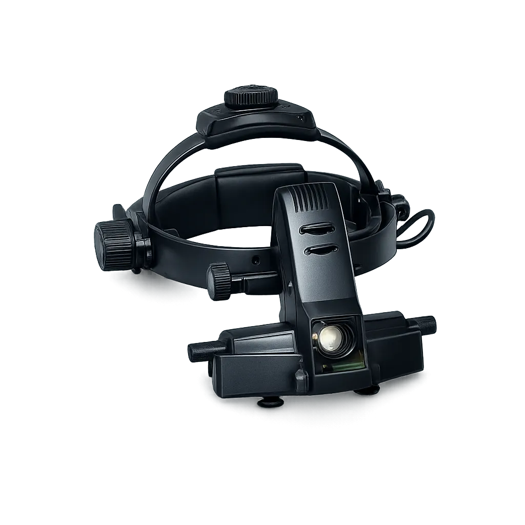
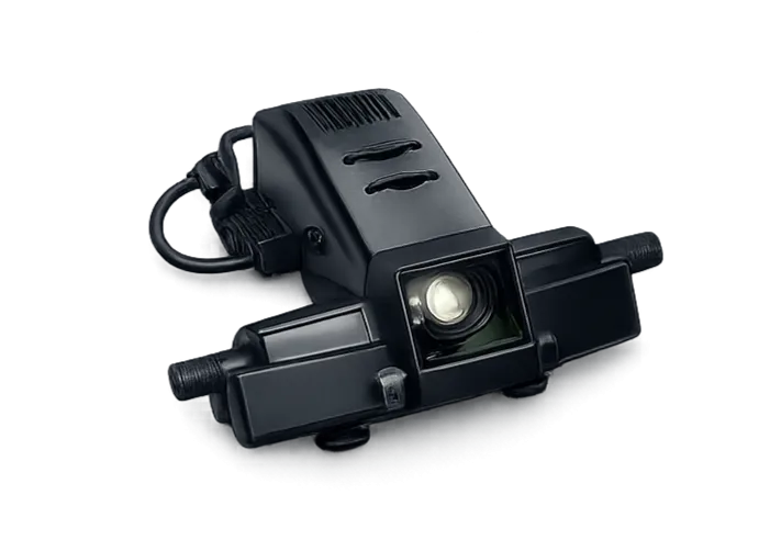
Indirect ophthalmoscopy gives a wide stereoscopic view of the fundus including the periphery, which direct ophthalmoscopy reaches poorly. What any examination reveals, and what follows from it, rests with the examining clinician rather than with the instrument producing the view. Direct ophthalmoscopy reaches the periphery poorly. Stereopsis assists that assessment. Elevation is appreciated rather than guessed at. Peripheral disease is reached properly.
Supervision and Training
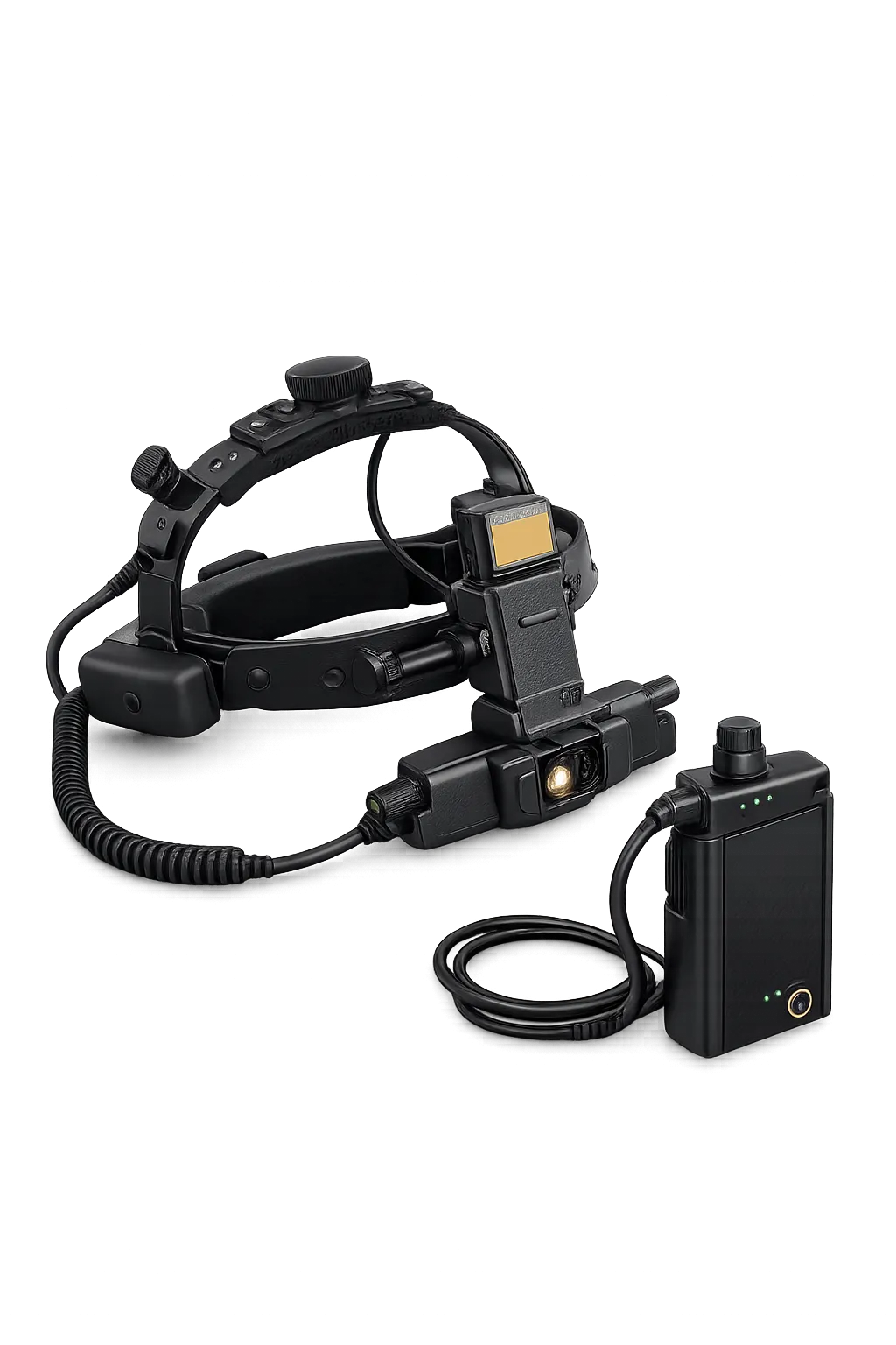
The teaching mirror allows a second person to observe the examiner's view directly, which is difficult to achieve any other way. For departments training junior staff, seeing what the examiner sees at the moment they see it is worth considerably more than describing it afterwards. Describing a finding afterwards is a poor substitute. Trainees see it as it happens. Seeing what the examiner sees is genuinely hard otherwise. Mirrors solve that directly.
Examination Rooms With Mains Access
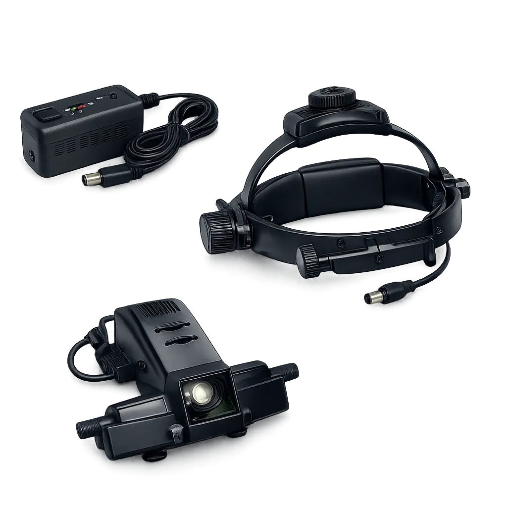
Because this model is mains powered, it suits a clinic room or theatre where a socket is available. Where examination must happen away from power, the cordless model in the same range answers that need instead. A socket must be available where it is used. The cordless model answers otherwise. Ward rounds are served poorly by a tether. Clinic rooms present no such difficulty.
Services With Decontamination Arrangements
The scleral depressor contacts the patient during examination, so it falls within a department's decontamination policy. What the depressor is made of and how it is cleaned should be established with Medigear.uk before the instrument enters service. Patient contact brings the depressor within policy. Material should be confirmed first. Decontamination policy governs any patient contact. Establishing it early avoids difficulty.