
Departments Already Performing Angiography
Fluorescein angiography requires trained staff, consent procedures and emergency provision that a service either has or does not. For departments already equipped for it, an instrument covering both colour imaging and angiography consolidates two functions; for those that are not, the equipment is the smaller part of what adopting angiography involves. Equipment is the smaller part of adopting the procedure. Consent procedures and emergency provision come first. Training follows behind those. A camera does not supply any of those requirements.
Routine Colour Fundus Imaging
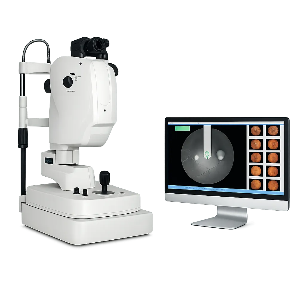
In its non-mydriatic mode the instrument images the retina through an undilated pupil, so ordinary colour work proceeds without drops or waiting. That mode carries none of the requirements angiography brings, and many appointments will use it alone. Throughput improves where drops are not required. Drops and waiting are removed from the appointment. Patients leave when imaging finishes. Many appointments will never need the other mode.
Patients Difficult to Position
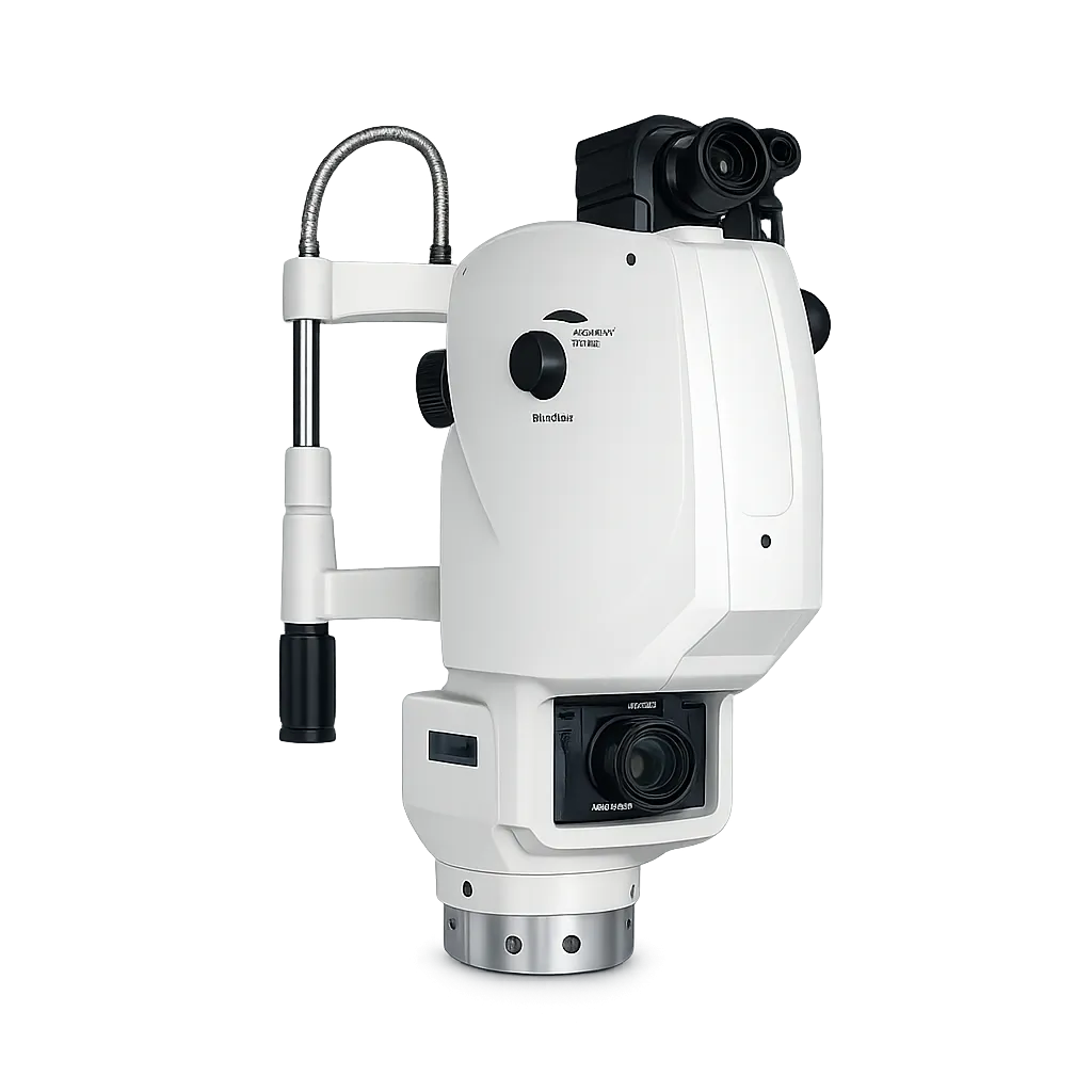
Head tilting through 30 degrees horizontally and 12.5 vertically, with a swinging optical body, extends the positions from which an eye can be reached. Where a patient cannot be moved easily, an instrument that moves instead is the practical answer. Frail patients benefit particularly from that reach. The instrument accommodates the patient rather than the reverse. Restricted mobility is common. Abandoned appointments waste a slot entirely.
Hospital Departments Needing Integration
DICOM 3.0 with HIS and PACS support means both colour and angiographic images join the patient record. For angiography in particular, where a sequence is reviewed against later studies, retrievability through the existing system matters considerably. Sequences are reviewed against later studies over time. Isolated archives make later comparison difficult. Integration is judged over years. Both image types join the same patient record.