
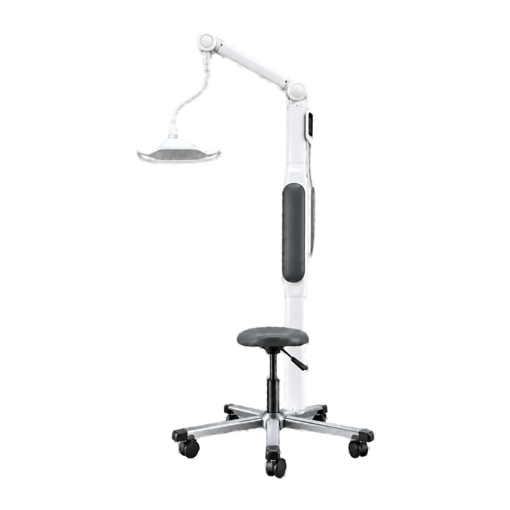
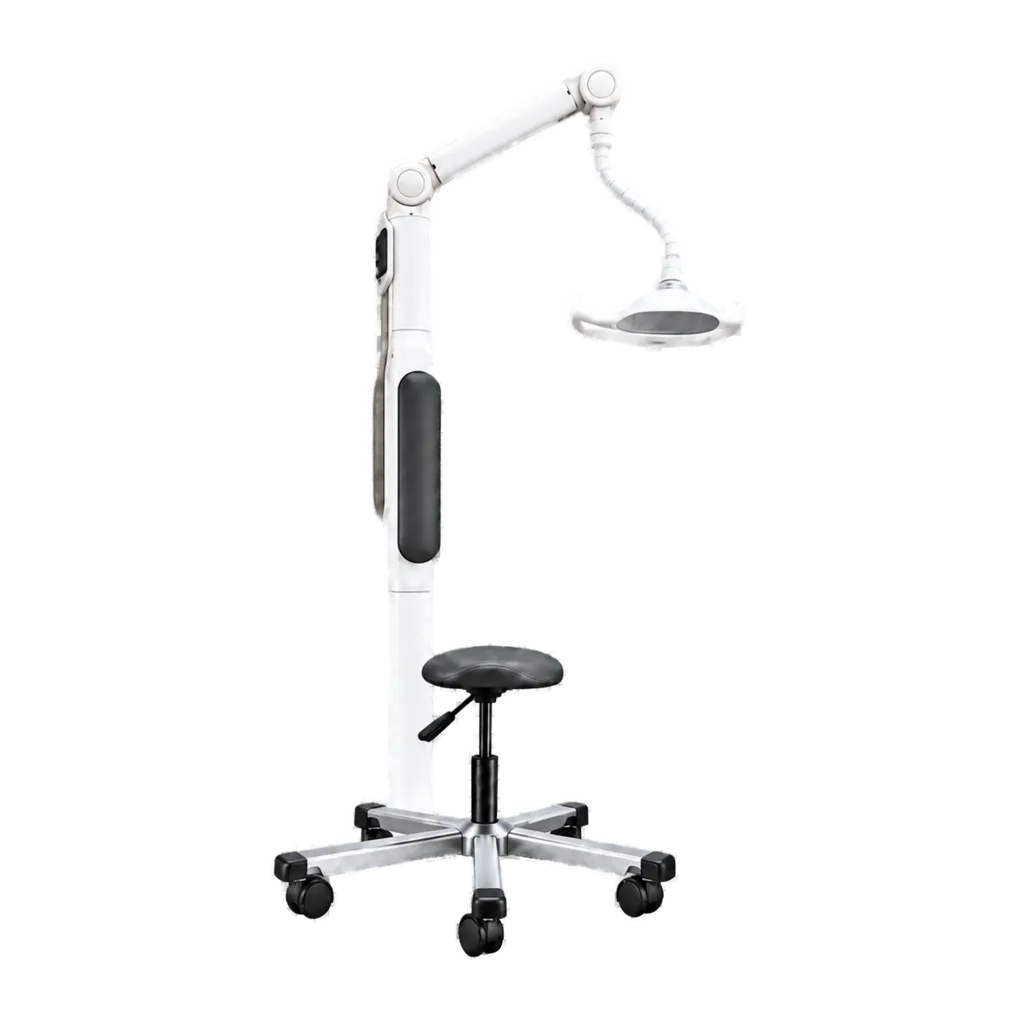
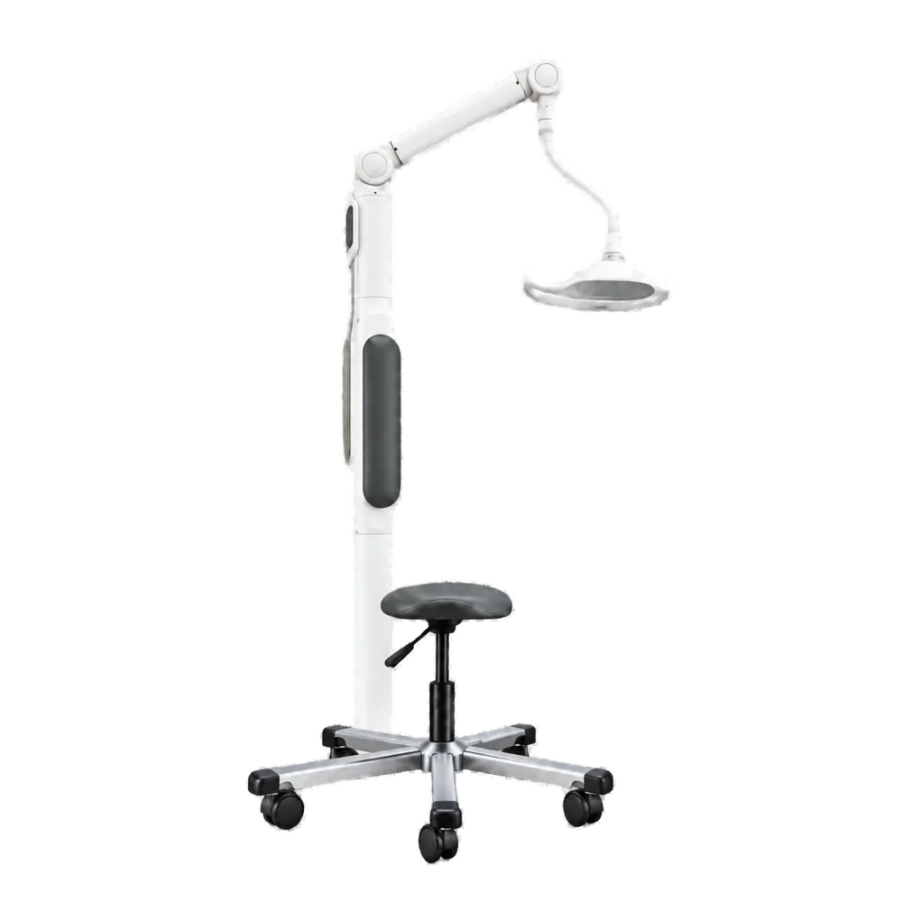
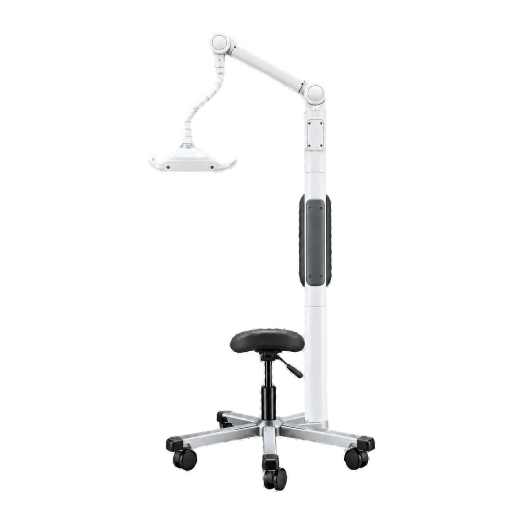
Intraoral radiography carries the diagnostic load in UK general dentistry, and this platform covers it across three installation types. It supports bitewing caries detection, periapical assessment of pathology, endodontic working-length and obturation verification, alveolar bone-level review, and root morphology assessment ahead of extraction.
Operation at 70 kV produces a longer greyscale, which suits views where crestal bone, root surface and restorative material must be read from one image, and reaches a given receptor density in a shorter exposure. An exposure floor of 0.06 seconds sits comfortably within the range direct digital sensors call for.
Throughput is where the duty cycle enters clinical planning. A practice taking occasional single radiographs will never notice it; one running full-mouth series or endodontic sequences will find it sets the pace of the appointment. Building the cooling interval into appointment lengths avoids an operator waiting at the chair with a patient in position.
Beam quality documentation supports commissioning. A published half-value layer gives the Radiation Protection Adviser a figure to check against rather than establish from scratch, which shortens the conversation at critical examination even though their own measurement governs.
Guidance from the College of General Dentistry and UKHSA Dental X-ray Protection Services remains the reference point UK practices work to, alongside HSE enforcement of IRR17.